

Review
doi: 10.1007/s12306-020-00650-2.
Epub 2020 Mar 3.
Assessment of the young adult hip joint using plain radiographs
Affiliations
- PMID: 32125641
- PMCID: PMC7686009
- DOI: 10.1007/s12306-020-00650-2
Item in Clipboard
Review
Assessment of the young adult hip joint using plain radiographs
Musculoskelet Surg.
2020 Dec.
Abstract
Radiographic examination remains the mainstay of the initial assessment of the young adult hip; however, common parameters are required to assist in the formation of accurate diagnoses and appropriate management plans. This paper aims to summarise the most important aspects of the assessment of plain radiographs performed on the young adult hip joint.
Keywords: Assessment; Hip joint; Radiographs; Young adult.
Conflict of interest statement
None.
Figures
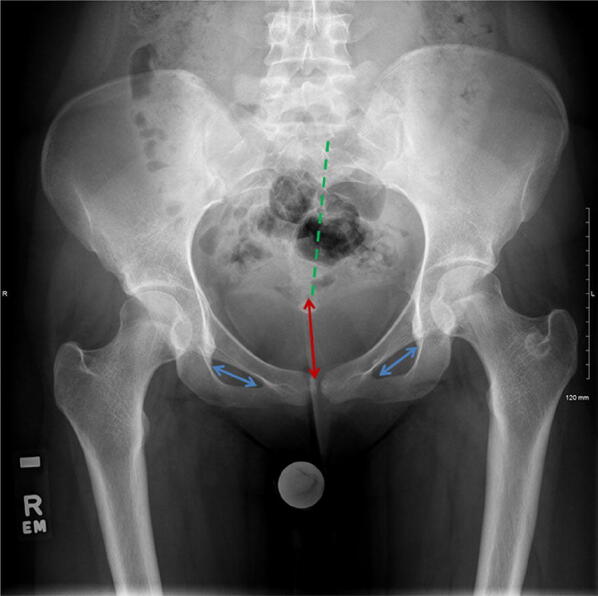
AP radiograph pelvis depicting pelvic rotation and tilt. The central sacral line (green dotted line) should be aligned centrally from the tip of the coccyx to the symphysis pubis. The obturator foramina (blue arrows), ischial spine and trochanters should be symmetrical. In this case, there is a slight pelvic rotation. The distance between sacro-coccygeal junction and the superior end of the symphysis (red arrow) is used to determine the pelvic tilt. A benign sclerotic ring is seen in the left trochanteric region
Cross-table lateral view
False profile lateral view
Frog lateral view

Dunn lateral view
a Teardrop sign. The U-shaped (blue line) teardrop consists of ilioischial line (red dotted arrow) and floor of the acetabulum. b Teardrop distance
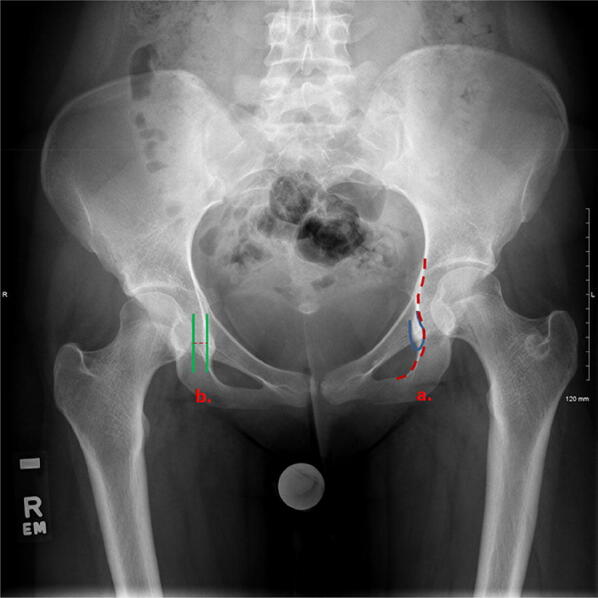
Sourcil (Tönnis) angle. The right hip (green angle) represents normal Sourcil angle, whereas the left hip (red angle) represents increased Sourcil angle. Lateral translation of the femoral head is clearly visible in keeping with hip dysplasia
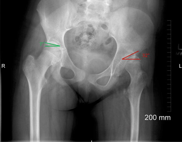
Anterior and posterior acetabular walls do not crossover and are only in contact at the lateral edge of the Sourcil suggestive of an anteverted acetabulum
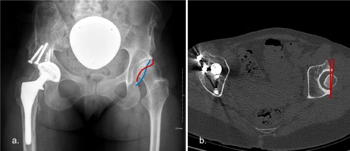
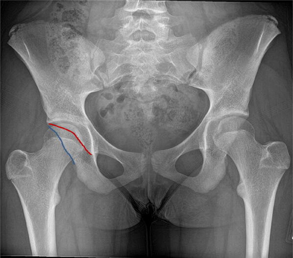
a AP radiograph pelvis. There is a crossover of the anterior and posterior walls of the left acetabulum, which represents acetabular retroversion. b CT axial image of the same patient which demonstrates left acetabular retroversion
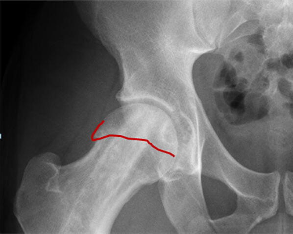
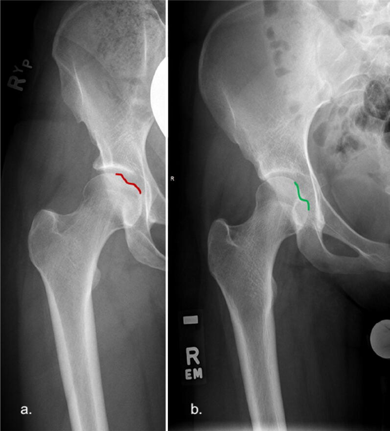
Red line represents a physeal scar which is seen to extend along the lateral margin of the femoral head–neck creating a CAM-type deformity
a Fovea on the right (red depression) is in contact with the Sourcil and represents Fovea alta. b Fovea on the left (green depression) is located medially and inferiorly and is therefore normally situated

Neck-shaft angle. a Right hip (red angle) measures 119° and therefore is regarded as coxa vara. b Left hip (green angle) measures 149° and therefore represents coxa valga
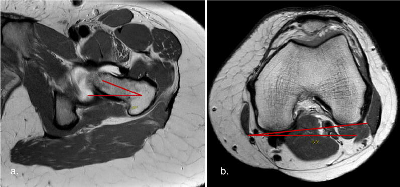
MR axial proton density (PD) sequences. a Level of the left femoral neck demonstrating femoral neck-horizontal angle. b Trans-condylar axis of the left knee demonstrating trans-condylar horizontal angle
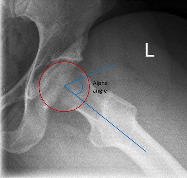
Alpha angle on lateral plain radiograph of left hip
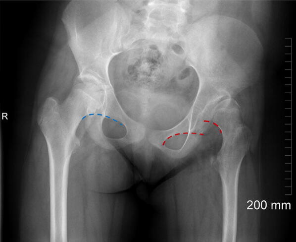
Right hip (blue dotted line) demonstrates preserved Shenton’s line. The left hip (red dotted line) represents ‘breaking’ of Shenton’s line resulting from superior and lateral subluxation of the dysplastic hip joint
References
Publication types
MeSH terms
LinkOut - more resources
Full Text Sources
