. 2020 Jun;41(6):1131-1137.
doi: 10.1002/humu.24008. Epub 2020 Mar 11.

Type II Alexander disease caused by splicing errors and aberrant overexpression of an uncharacterized GFAP isoform

Affiliations

Type II Alexander disease caused by splicing errors and aberrant overexpression of an uncharacterized GFAP isoform

Guy Helman et al. Hum Mutat. 2020 Jun.

Erratum in

Abstract

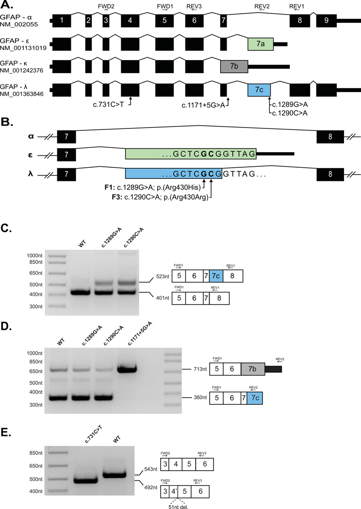
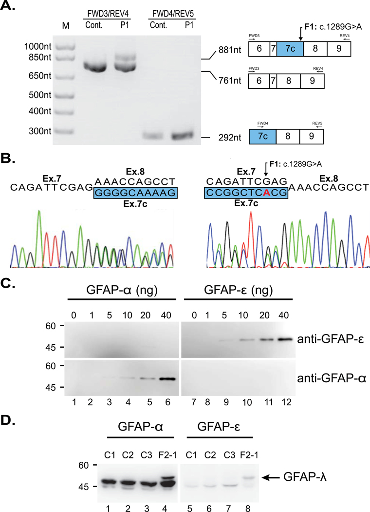
Alexander disease results from gain-of-function mutations in the gene encoding glial fibrillary acidic protein (GFAP). At least eight GFAP isoforms have been described, however, the predominant alpha isoform accounts for ∼90% of GFAP protein. We describe exonic variants identified in three unrelated families with Type II Alexander disease that alter the splicing of GFAP pre-messenger RNA (mRNA) and result in the upregulation of a previously uncharacterized GFAP lambda isoform (NM_001363846.1). Affected members of Family 1 and Family 2 shared the same missense variant, NM_001363846.1:c.1289G>A;p.(Arg430His) while in Family 3 we identified a synonymous variant in the adjacent nucleotide, NM_001363846.1:c.1290C>A;p.(Arg430Arg). Using RNA and protein analysis of brain autopsy samples, and a mini-gene splicing reporter assay, we demonstrate both variants result in the upregulation of the lambda isoform. Our approach demonstrates the importance of characterizing the effect of GFAP variants on mRNA splicing to inform future pathophysiologic and therapeutic study for Alexander disease.

Keywords: Alexander disease; aberrant splicing; leukodystrophy.


Figures

Figure 1:
Figure 1:. Missense and synonymous variants in GFAP result in alteration in GFAP pre-mRNA splicing.
(A) Schematic of four GFAP mRNA isoforms. Exons 1–9 are numbered per the GFAP-α isoform. Introns are not shown to scale. (B) Schematic of C-terminal variants between exons 7–8 of GFAP showing the positions of the single nucleotide substitutions identified in Family 1 (F1) and Family 3 (F3) relative to exon 7A (green, GFAP-ε) and exon 7C (blue, GFAP-λ). (C) A GFAP mini-gene plasmid was created by introducing 8.1kb of GFAP genomic DNA (exon1 – exon 9, amino acids 57–433) into the mammalian expression vector pEYFP-C1 (see Methods). Versions of this construct were generated containing the GFAP-ε variants, c.1289G>A and c.1290C>A, or the GFAP-α variants c.731C>T and c.1171+5G>A variants before transfection into HEK cells. rtPCR amplification from exon 5 to exon 8 yielded a predominant amplicon consistent with the GFAP-α isoform (401nt) from the WT (Lane 1) c.1289G>A (Lane 2) and c.1290C>A (Lane 3) constructs. The c.1289G>A and c.1290C>A mutant constructs also generated a larger amplicon consistent with the GFAP-λ isoform (523nt). (D) Amplification from exon 5 to exon 7A/7C yielded a predominant amplicon consistent with the GFAP-ε and GFAP-λ isoforms (360nt) from the WT (Lane 1), c.1289G>A (Lane 2) and c.1290C>A (Lane 3) constructs, and a minor amplicon consistent with the GFAP-κ (713nt) is also visible in each of these lanes. The c.1171+5G>A mutant construct yielded a single predominant amplicon consistent in size with GFAP-κ (713nt). (E) Amplification from exon 3 to exon 5 (common to all major GFAP isoforms) yielded a single amplicon consistent with the expected size of 543nt from the WT construct (Lane 2). Amplification from the construct containing the c.731C>T missense variant (Lane 1) yielded a smaller single amplicon consistent with a truncation of exon 4. Sanger sequencing of the c.731C>T amplicon confirmed a 51nt truncation of exon 4 (Supp. Figure S3).
Figure 2:
Figure 2:. Demonstration of the presence of GFAP-λ transcript and protein in the AxD brain.
(A) RT-PCR of mRNA from autopsy brain tissue of an affected member of Family 1 (F1-1) and an unaffected control. Lanes 1 and 2: amplification between exons 6 and 9 generates an amplicon of the expected 761nt from both the patient and control cDNA samples. An additional 881nt amplicon can be seen in the cDNA sample of the affected individual (F1-1), consistent with GFAP-λ. Lanes 3 and 4: amplification between exons 7C and 9 generates a GFAP-λ specific amplicon of 292nt that is detectible in the control cDNA sample but noticeably more abundant when amplified from the patient cDNA. (B) Sanger sequencing of the mixed amplicon from lane 2 in both forward (left) and reverse (right; reverse complement shown) shows the expected nucleotide sequences and splice sites for GFAP-α and GFAP-λ. (C) The specificity and affinity of the isoform-specific GFAP antibodies were characterized by immunoblotting using dilutions of known concentrations of purified recombinant human GFAP-α (lanes 1-6) and GFAP-ε (lanes 7-12). The anti-GFAP-α and anti-GFAP-ε antibodies recognized purified GFAP-α (lanes 4-6, bottom panel) and GFAP-ε (lanes 9-12, top panel), respectively, confirming the specificity of these antibodies. (D) Intermediate filament enriched fractions were prepared from brains of four AxD affected individuals (lanes 1-4), including an affected family member of Family 2 (F2-1, lane 4). Samples (10-12 μg protein load per lane) were analyzed by immunoblotting with anti-GFAP-α (lanes 1-4) and anti-GFAP-ε (lanes 5-8) antibodies. Note the presence of GFAP-λ in F2-1 (lanes 4 and 8, arrow).

References

    1. Blechingberg J, Holm IE, Nielsen KB, Jensen TH, Jørgensen AL, & Nielsen AL (2007). Identification and characterization of GFAPκ, a novel glial fibrillary acidic protein isoform. Glia, 55(5), 497–507. doi:10.1002/glia.20475 - DOI - PubMed
    1. Bugiani M, Boor I, van Kollenburg B, Postma N, Polder E, van Berkel C, . . . van der Knaap MS (2011). Defective Glial Maturation in Vanishing White Matter Disease. Journal of Neuropathology & Experimental Neurology, 70(1), 69–82. doi:10.1097/NEN.0b013e318203ae74 - DOI - PMC - PubMed
    1. Flint D, Li R, Webster LS, Naidu S, Kolodny E, Percy A, . . . Brenner M (2012). Splice site, frameshift, and chimeric GFAP mutations in Alexander disease. Hum Mutat, 33(7), 1141–1148. doi:10.1002/humu.22094 - DOI - PMC - PubMed
    1. Graff-Radford J, Schwartz K, Gavrilova RH, Lachance DH, & Kumar N (2014). Neuroimaging and clinical features in type II (late-onset) Alexander disease. Neurology, 82(1), 49–56. doi:10.1212/01.wnl.0000438230.33223.bc - DOI - PMC - PubMed
    1. Green L, Berry IR, Childs A-M, McCullagh H, Jose S, Warren D, . . . Livingston JH (2018). Whole Exon Deletion in the GFAP Gene Is a Novel Molecular Mechanism Causing Alexander Disease. Neuropediatrics, 49(02), 118–122. doi:10.1055/s-0037-1608921 - DOI - PubMed

Publication types