Review
. 2020 Mar 3;15(1):20.
doi: 10.1186/s13000-020-00943-x.

Fibromatosis-like metaplastic carcinoma: a case report and review of the literature

Affiliations
Review

Fibromatosis-like metaplastic carcinoma: a case report and review of the literature

Jasper Victoor et al. Diagn Pathol. .

Abstract

Background: We report an unusual case of low-grade fibromatosis-like metaplastic carcinoma (LG-FLMC) of the breast. This exceedingly rare epithelial breast malignancy has been reported only 68 times in the past 20 years, and is classified as a subtype of metaplastic breast carcinoma (MBC). It is a locally aggressive tumor with a low potential for lymph node and distant metastases, but with a tendency to recur after excision. Here we describe a less common presentation of LG-FLMC, provide its molecular characterization, discuss the major differential diagnosis and bring a short review of the literature.

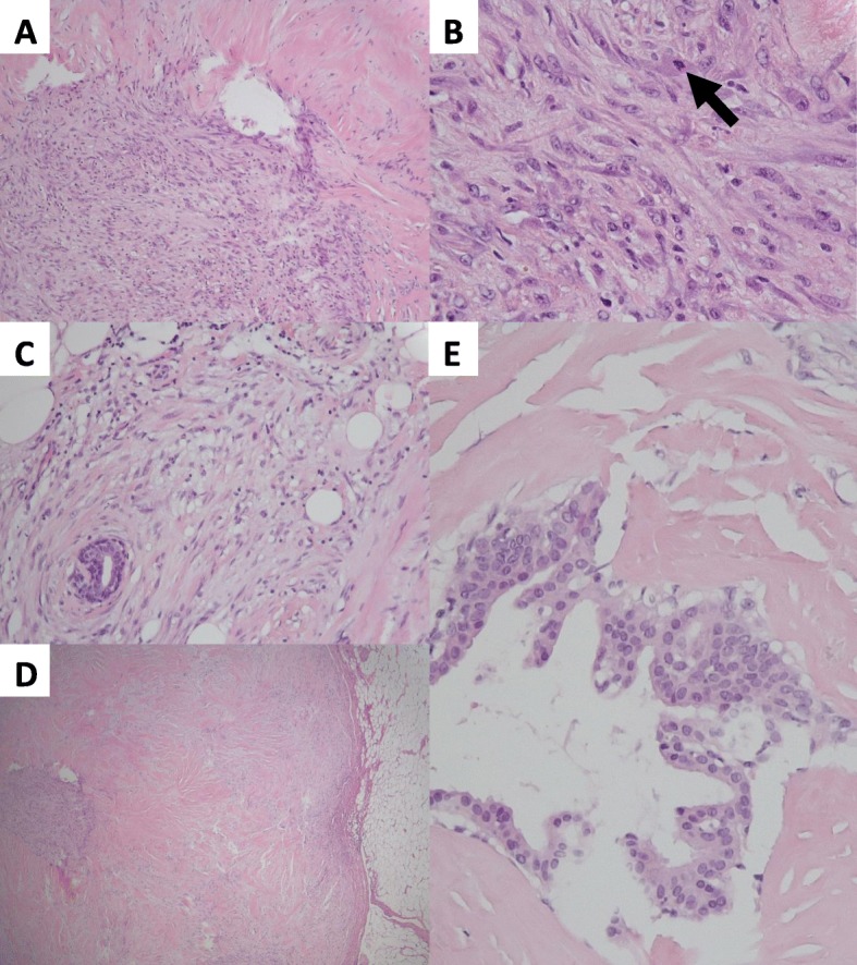
Case presentation: A 65-year-old woman presented with a self-palpated breast lump that had discordant radio-pathological features. While imaging results were compatible with an infiltrative malignancy, on core needle biopsy (CNB) a sharply delineated lesion composed by a bland-looking population of spindle cells was observed; excision was recommended for final diagnosis. Histology of the resection specimen showed small areas of epithelial differentiation and foci of peripheral invasion. Immunohistochemical analysis revealed a co-immunoreactivity for epithelial and myoepithelial markers in the spindle cell component. Mutation analysis with a capture-based next generation sequencing method revealed pathogenic mutations in GNAS, TERT-promotor and PIK3R1 genes. A diagnosis of LG-FLMC was rendered.

Conclusion: This case highlights the importance of a broad differential diagnosis, exhaustive sampling and the use of a broad immunohistochemical panel whenever dealing with a low-grade spindle cell lesion in the breast, and provides further insights into the molecular background of LG-FLMC.

Keywords: Breast; Low-grade fibromatosis-like metaplastic carcinoma; Metaplastic breast carcinoma; Spindle cell lesion.


Conflict of interest statement

The authors declare that they have no competing interests.

Figures

Fig. 1
Fig. 1
Routine histology with hematoxylin and eosin (H&E) stains. a The breast lesion has a variable cellularity. Large areas consist of dense collagenous stroma with a low cellularity (upper right of the picture). These areas are admixed with more cellular areas that have a myxoid appearance (lower left of the picture). (H&E ×50). b A highly cellular, myxoid area of the lesion. There is clustering of fusiform to discrete epithelioid cells with a round, slightly irregular nucleus that contains a small nucleolus. Occasional mitotic figures are seen (arrow). (H&E × 400). c Within the breast lesion, small and normal-looking ducts are entrapped by the proliferative spindle cells. The latter show no obvious atypia or mitotic activity at high magnification. At the periphery, a limited infiltration of bland-looking spindle cells into the surrounding fat tissue is seen. A scattered infiltrate of lymphocytes is also seen. (H&E × 100). d A view of the assessable border of the breast lesion at low magnification. A nodular and sharply delineated margin in relation to the surrounding fat tissue can be seen. This is not typical for FLMC, where a more infiltrative growth pattern is expected at the border. (H&E × 25). e In the center of the lesion, a distinctive area with a relatively high number of bland-looking ducts surrounded by a striking sclerotic stroma is seen. (H&E × 200)
Fig. 2
Fig. 2
Immunohistochemistry. Staining patterns for αSMA (a), p63 (b), CK AE1/AE3 (c) and β-catenin (d). (IHC × 400). There is a co-expression in both the spindle cells and the more epithelioid cells for p63 and CK AE1/AE3

References

    1. Rakha EA, Aleskandarany MA, Lee AHS, Ellis IO. An approach to the diagnosis of spindle cell lesions of the breast. Histopathology. 2016;68:33–44. doi: 10.1111/his.12865. - DOI - PubMed
    1. Tay TKY, Tan PH. Spindle cell lesions of the breast - an approach to diagnosis. Semin Diagn Pathol. 2017;34:400–409. doi: 10.1053/j.semdp.2017.05.012. - DOI - PubMed
    1. Geyer FC, Pareja F, Weigelt B, Rakha E, Ellis IO, Schnitt SJ, Reis-Filho JS. The spectrum of triple-negative breast disease: high- and low-grade lesions. Am J Pathol. 2017;187:2139–2151. doi: 10.1016/j.ajpath.2017.03.016. - DOI - PMC - PubMed
    1. Nonnis R, Paliogiannis P, Giangrande D, Marras V, Trignano M. Low-grade fibromatosis-like spindle cell metaplastic carcinoma of the breast: a case report and literature review. Clin Breast Cancer. 2012;12:147–150. doi: 10.1016/j.clbc.2012.01.011. - DOI - PubMed
    1. Lakhani SR, Ellis IO, Schnitt SJ, Tan PH, van de Vijver MJ. WHO classification of tumours of the breast. 4. Lyon: IARC; 2012. pp. 48–52.

Substances

LinkOut - more resources