Review
. 2020 May;19(5):757-773.
doi: 10.1074/mcp.R120.001941. Epub 2020 Mar 3.

Proximity Dependent Biotinylation: Key Enzymes and Adaptation to Proteomics Approaches

Affiliations
Review

Proximity Dependent Biotinylation: Key Enzymes and Adaptation to Proteomics Approaches

Payman Samavarchi-Tehrani et al. Mol Cell Proteomics. 2020 May.

Abstract

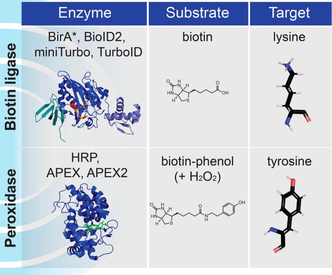
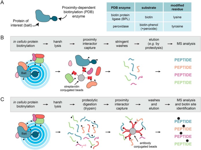
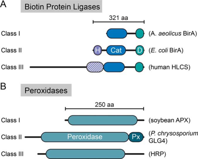
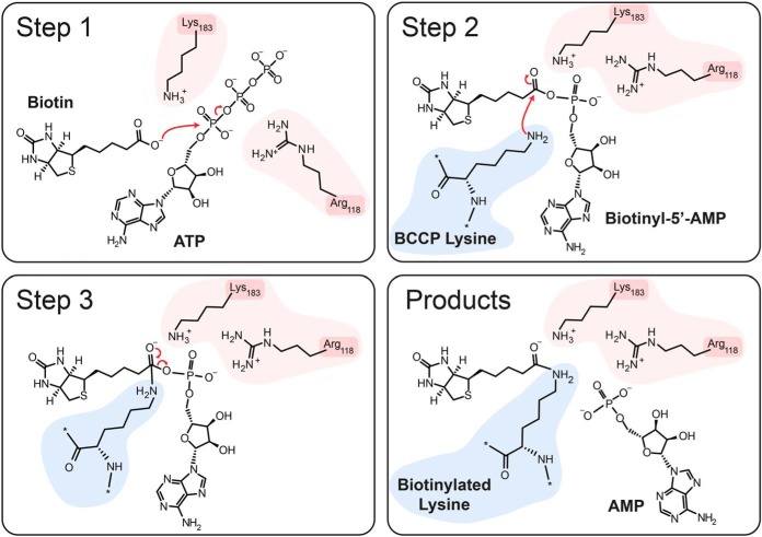
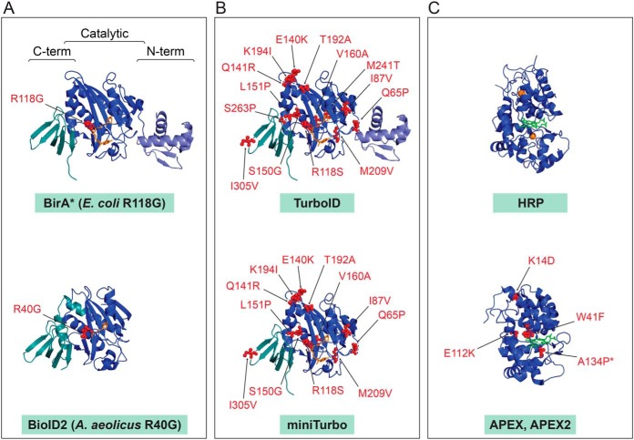
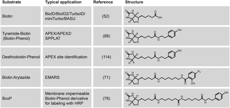
The study of protein subcellular distribution, their assembly into complexes and the set of proteins with which they interact with is essential to our understanding of fundamental biological processes. Complementary to traditional assays, proximity-dependent biotinylation (PDB) approaches coupled with mass spectrometry (such as BioID or APEX) have emerged as powerful techniques to study proximal protein interactions and the subcellular proteome in the context of living cells and organisms. Since their introduction in 2012, PDB approaches have been used in an increasing number of studies and the enzymes themselves have been subjected to intensive optimization. How these enzymes have been optimized and considerations for their use in proteomics experiments are important questions. Here, we review the structural diversity and mechanisms of the two main classes of PDB enzymes: the biotin protein ligases (BioID) and the peroxidases (APEX). We describe the engineering of these enzymes for PDB and review emerging applications, including the development of PDB for coincidence detection (split-PDB). Lastly, we briefly review enzyme selection and experimental design guidelines and reflect on the labeling chemistries and their implication for data interpretation.

Keywords: APEX; BioID; Protein-protein interactions; biotin ligase; cellular organelles; enzymes; mass spectrometry; molecular biology; peroxidase; protein engineering; proximity-dependent biotinylation.


Conflict of interest statement

The authors declare that they have no conflicts of interest with the contents of this article

Figures

None
Graphical abstract
Fig. 1.
Fig. 1.
Principles of Proximity-Dependent Biotinylation. A, A protein of interest (bait) is fused in-frame to a PDB enzyme from one of two families, biotin protein ligases or peroxidases, that have distinct substrate requirements and modify different amino acids. B, Schematic workflow for a PDB experiment identifying proximal proteins. Proteins are labeled inside living cells prior to a harsh lysis and a protein-level capture on streptavidin beads. After stringent washing, elution is most often effected by proteolysis with trypsin, and the non-biotinylated peptides are released and identified by mass spectrometry. A variation of this approach consists of performing an elution with high concentrations of acid such as trifluoroacetic acid; in this case information about the site of biotinylation may be obtained. C, Alternative workflow for peptide-level capture and identification of the biotinylated peptides: an antibody against biotin is used to capture biotinylated peptides directly. Alternatively, other biotin affinity capture strategies can be employed.
Fig. 2.
Fig. 2.
Domain structure of the different classes of PDB enzymes. A, Biotin protein ligases. H: helix-turn-helix DNA binding domain; Cat: central catalytic domain (in blue); D: domain of unknown function. Examples of proteins for each of the three structural classes are shown. B, Peroxidases. APX: Ascorbate peroxidase; GLG4: P. chrysosporium Ligninase H2; HRP: Horseradish peroxidase. Px: peroxidase extension region, peroxidase catalytic domain (in teal).
Fig. 3.
Fig. 3.
Mechanism of labeling by BPLs. Side chains on the BPL enzyme involved in the reaction are highlighted in pink, whereas the lysine side chain of the substrate (here BCCP) is shown in blue. See text for details.
Fig. 4.
Fig. 4.
Structures of the main enzymes employed in PDB research. PDB files were downloaded from the RCSB Protein Data Bank and visualized in Pymol. Mutations over the wild type enzymes are indicated. A, Original enzymes used in BioID. Structures of the wild type E. coli BirA with biotinyl-5′-AMP (source PDB 2EWN) and A. aeolicus BPL with biotin (source PDB 3EFS) with position of the mutation decreases the affinity for biotinyl-5′-AMP intermediate. B, Molecularly evolved TurboID and miniTurbo displayed on the E. coli wild type BirA structure. C, Peroxidases. HRP with heme and calcium ions (source 1HCH) and APEX/APEX2 mutations displayed on the structure of soybean APX with heme (source PDB 1OAG) are indicated (note that APEX2 contains the additional A134P mutation compared with APEX).
Fig. 5.
Fig. 5.
Substrates used in PDB approaches. Different related substrates for PDB have been developed specifically for different variations on the PDB theme. See text and references for details.

References

    1. Yates J. R. 3rd, Gilchrist A., Howell K. E., and Bergeron J. J. (2005) Proteomics of organelles and large cellular structures. Nat. Rev. 6, 702–714 - PubMed
    1. Itzhak D. N., Tyanova S., Cox J., and Borner G. H. (2016) Global, quantitative and dynamic mapping of protein subcellular localization. eLife 5 - PMC - PubMed
    1. Mulvey C. M., Breckels L. M., Geladaki A., Britovsek N. K., Nightingale D. J. H., Christoforou A., Elzek M., Deery M. J., Gatto L., and Lilley K. S. (2017) Using hyperLOPIT to perform high-resolution mapping of the spatial proteome. Nat. Protocols 12, 1110–1135 - PubMed
    1. Geladaki A., Kocevar Britovsek N., Breckels L. M., Smith T. S., Vennard O. L., Mulvey C. M., Crook O. M., Gatto L., and Lilley K. S. (2019) Combining LOPIT with differential ultracentrifugation for high-resolution spatial proteomics. Nat. Commun. 10, 331. - PMC - PubMed
    1. Kristensen A. R., Gsponer J., and Foster L. J. (2012) A high-throughput approach for measuring temporal changes in the interactome. Nat. Methods 9, 907–909 - PMC - PubMed

Publication types

Grants and funding