

Accumulation of Globotriaosylceramide in Podocytes in Fabry Nephropathy Is Associated with Progressive Podocyte Loss
- PMID: 32127409
- PMCID: PMC7191924
- DOI: 10.1681/ASN.2019050497
Accumulation of Globotriaosylceramide in Podocytes in Fabry Nephropathy Is Associated with Progressive Podocyte Loss
Abstract
Background: In males with classic Fabry disease, the processes leading to the frequent outcome of ESKD are poorly understood. Defects in the gene encoding α-galactosidase A lead to accumulation of globotriaosylceramide (GL3) in various cell types. In the glomerular podocytes, accumulation of GL3 progresses with age. Of concern, podocytes are relatively resistant to enzyme replacement therapy and are poorly replicating, with little ability to compensate for cell loss.
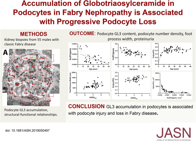
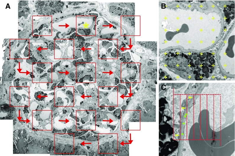
Methods: In this study of 55 males (mean age 27 years) with classic Fabry disease genotype and/or phenotype, we performed unbiased quantitative morphometric electron microscopic studies of biopsied kidney samples from patients and seven living transplant donors (to serve as controls). We extracted clinical information from medical records and clinical trial databases.
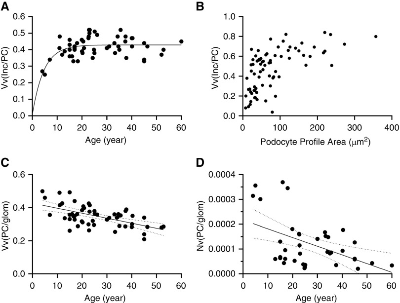
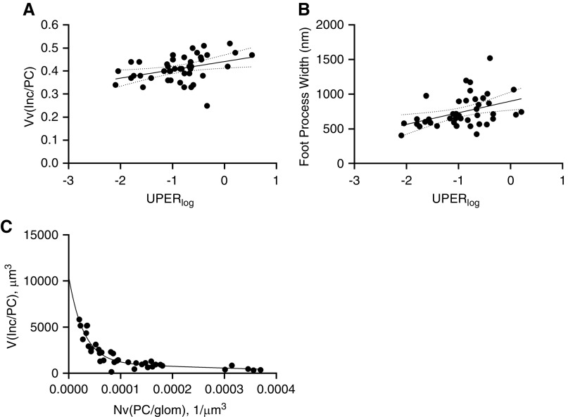
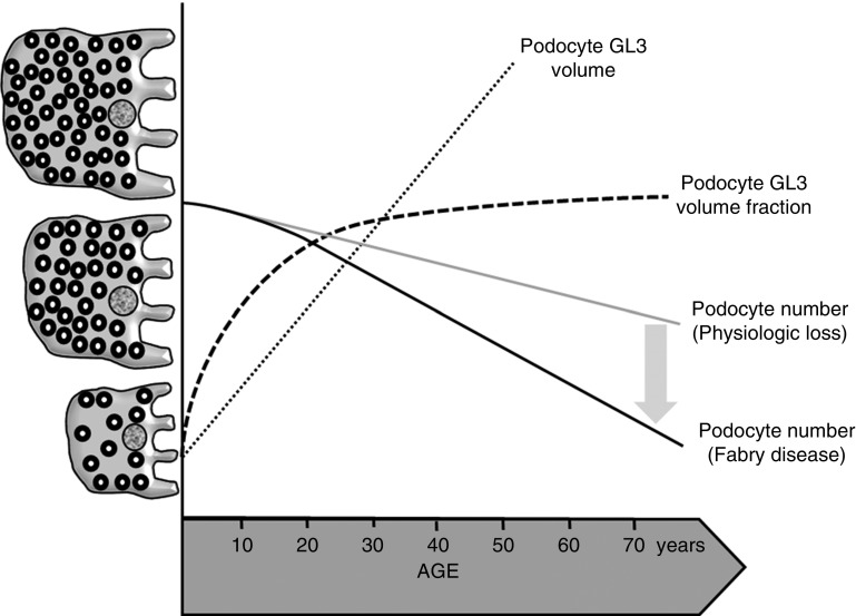
Results: Podocyte GL3 volume fraction (proportion of podocyte cytoplasm occupied by GL3) increased with age up to about age 27, suggesting that increasing podocyte GL3 volume fraction beyond a threshold may compromise survival of these cells. GL3 accumulation was associated with podocyte injury and loss, as evidenced by increased foot process width (a generally accepted structural marker of podocyte stress and injury) and with decreased podocyte number density per glomerular volume. Worsening podocyte structural parameters (increasing podocyte GL3 volume fraction and foot process width) was also associated with increasing urinary protein excretion-a strong prognosticator of adverse renal outcomes in Fabry disease-as well as with decreasing GFR.
Conclusions: Given the known association between podocyte loss and irreversible FSGS and global glomerulosclerosis, this study points to an important role for podocyte injury and loss in the progression of Fabry nephropathy and indicates a need for therapeutic intervention before critical podocyte loss occurs.
Keywords: Fabry disease; chronic kidney disease; pathology; podocyte.
Copyright © 2020 by the American Society of Nephrology.
Figures
References
-
- Desnick RJ, Allen KY, Desnick SJ, Raman MK, Bernlohr RW, Krivit W: Fabry’s disease: Enzymatic diagnosis of hemizygotes and heterozygotes. Alpha-galactosidase activities in plasma, serum, urine, and leukocytes. J Lab Clin Med 81: 157–171, 1973 - PubMed
-
- Thurberg BL, Rennke H, Colvin RB, Dikman S, Gordon RE, Collins AB, et al.: Globotriaosylceramide accumulation in the Fabry kidney is cleared from multiple cell types after enzyme replacement therapy. Kidney Int 62: 1933–1946, 2002 - PubMed
-
- Germain DP, Waldek S, Banikazemi M, Bushinsky DA, Charrow J, Desnick RJ, et al.: Sustained, long-term renal stabilization after 54 months of agalsidase beta therapy in patients with Fabry disease. J Am Soc Nephrol 18: 1547–1557, 2007 - PubMed
-
- Banikazemi M, Bultas J, Waldek S, Wilcox WR, Whitley CB, McDonald M, et al.; Fabry Disease Clinical Trial Study Group: Agalsidase-beta therapy for advanced Fabry disease: A randomized trial. Ann Intern Med 146: 77–86, 2007 - PubMed
Publication types
MeSH terms
Substances
Grants and funding
LinkOut - more resources
Full Text Sources
Medical
