. 2020 Mar 3;11(2):e00027-20.
doi: 10.1128/mBio.00027-20.

Heterosubtypic Protection Induced by a Live Attenuated Influenza Virus Vaccine Expressing Galactose-α-1,3-Galactose Epitopes in Infected Cells

Affiliations

Heterosubtypic Protection Induced by a Live Attenuated Influenza Virus Vaccine Expressing Galactose-α-1,3-Galactose Epitopes in Infected Cells

Li-Meng Yan et al. mBio. .

Abstract

Anti-galactose-α-1,3-galactose (anti-α-Gal) antibody is naturally expressed at a high level in humans. It constitutes about 1% of immunoglobulins found in human blood. Here, we designed a live attenuated influenza virus vaccine that can generate α-Gal epitopes in infected cells in order to facilitate opsonization of infected cells, thereby enhancing vaccine-induced immune responses. In the presence of normal human sera, cells infected with this mutant can enhance phagocytosis of human macrophages and cytotoxicity of NK cells in vitro Using a knockout mouse strain that allows expression of anti-α-Gal antibody in vivo, we showed that this strategy can increase vaccine immunogenicity and the breadth of protection. This vaccine can induce 100% protection against a lethal heterosubtypic group 1 (H5) or group 2 (mouse-adapted H3) influenza virus challenge in the mouse model. In contrast, its heterosubtypic protective effect in wild-type or knockout mice that do not have anti-α-Gal antibody expression is only partial, demonstrating that the enhanced vaccine-induced protection requires anti-α-Gal antibody upon vaccination. Anti-α-Gal-expressing knockout mice immunized with this vaccine produce robust humoral and cell-mediated responses upon a lethal virus challenge. This vaccine can stimulate CD11blo/- pulmonary dendritic cells, which are known to be crucial for clearance of influenza virus. Our approach provides a novel strategy for developing next-generation influenza virus vaccines.IMPORTANCE Influenza A viruses have multiple HA subtypes that are antigenically diverse. Classical influenza virus vaccines are subtype specific, and they cannot induce satisfactory heterosubtypic immunity against multiple influenza virus subtypes. Here, we developed a live attenuated H1N1 influenza virus vaccine that allows the expression of α-Gal epitopes by infected cells. Anti-α-Gal antibody is naturally produced by humans. In the presence of this antibody, human cells infected with this experimental vaccine virus can enhance several antibody-mediated immune responses in vitro Importantly, mice expressing anti-α-Gal antibody in vivo can be fully protected by this H1N1 vaccine against a lethal H5 or H3 virus challenge. Our work demonstrates a new strategy for using a single influenza virus strain to induce broadly cross-reactive immune responses against different influenza virus subtypes.

Keywords: immunology; influenza; influenza virus vaccines; live vector vaccines; universal vaccine.


Figures

FIG 1
FIG 1
Characterization of the NAGT mutant in vitro. (A) WT and mutated NA segments in the cRNA sense strand. A mouse α-1,3-GT gene ORF was introduced immediately before the stop codon of the NA ORF (open arrow). An autoproteolytic cleavage site of porcine teschovirus-1 flanked by two short peptide linkers (GSG) was introduced into the junction of NA and α-1,3-GT ORF as shown. 5′ and 3′ untranslated regions (UTRs) and the start codon (black arrow) are indicated. The red box represents the 5′-end vRNA packaging signal sequence at the NA ORF region. (B) Detection of influenza virus NA protein infection by Western blotting. Human A549 cells infected with WT and NAGT viruses (MOI, 1) were harvested at 48 h postinfection. (C) Replication kinetics of NAGT mutant in MDCK cells (MOI, 0.001). Progeny virus titers were determined using standard plaque assays. (D) Immunofluorescent staining of α-Gal epitopes in human A549 cells infected with WT or NAGT virus at 24 h postinfection (MOI, 1). Signals from the nuclear counterstain (DAPI) are also shown. (E) Human A549 cells infected with the NAGT virus can enhance phagocytic activities of human monocyte-derived macrophages. (F) Antibody-dependent NK cell assay. Human A549 cells treated with PBS (left), WT virus (middle), or the NAGT virus (right) were incubated with activated human NK cells in the presence (top) or absence (bottom) of heat-inactivated human serum. The percentages of influenza virus NP-positive cells after incubation under different experimental conditions are shown. These images show representative fluorescence-activated cell sorting (FACS) plots obtained by using a serum sample from a healthy donor. (G) Stimulating effect of all tested human sera on NK cells for killing WT- and NAGT-infected A549 cells (fold reduction in NP+ A549 cells; n = 6; paired t test). (H) Luciferase reporter assay for ADCC activity. A549 cells infected with the WT or NAGT virus were first treated with serially diluted human serum samples and then tested by ADCC assays. Each data point represents the average reading from six different human serum samples; values are means ± standard deviations (SD). *, P < 0.05; **, P < 0.01; ***, P < 0.001.
FIG 2
FIG 2
Vaccination with the NAGT mutant in WT and α-1,3-GT KO mice. (A) General scheme of vaccination and virus challenge. Mice were primed with rabbit RBCs and given boosters via the intraperitoneal route at 4 and 6 weeks of age. Mice were vaccinated with the NAGT mutant at 8 weeks of age and then challenged with a lethal dose of influenza virus via the intranasal route. Relevant samples were collected at 4, 6, and 8 weeks of age and at various postvaccination (days 1, 5, and 11) and postchallenge (days 0, 3, and 7) time points. (B) Anti-α-Gal antibody titers in different experimental groups before virus challenge in week 11. (C) H1N1 and H3N2 virus-specific neutralizing antibody titers in different experimental groups before virus challenge in week 11. (D and E) Splenic CD4+ and CD8+ T-cell recall responses against different influenza viruses (H1N1/PR8, H3N2/HK68, H1N1/Brisbane/07, and H5N2/HK/MPF461/07). T cells were studied with ICS assays. Percentages of activated T cells (TNF-α+) are shown. Data are means ± SD. ***, P < 0.001.
FIG 3
FIG 3
Professional antigen-presenting cells (DCs and macrophages) and neutrophils in spleen tissue after vaccination. Spleens from vaccinated WT and KO mice were harvested at days 1, 5, and 21 postvaccination. Unvaccinated mice (−) were used as controls. Representative FACS plots and percentages of DCs (A), neutrophils (B), and macrophages (C) in all studied samples are shown. Data from vaccinated mice are highlighted. The dotted line shows the comparison between KO and WT mice at the same time point (t test). Data are means ± SD. *, P < 0.05; **, P < 0.01.
FIG 4
FIG 4
Professional antigen-presenting cells (DCs and alveolar macrophages) and neutrophils in lung tissue after vaccination. Lungs from vaccinated WT and KO mice were harvested at days 1, 5, and 21 postvaccination. Unvaccinated mice (-) were used as controls. Representative FACS plots and percentages of DCs (A), CD11blo/− and CD11b+ DCs (B), neutrophils (C), and alveolar macrophages (D) in all studied samples are shown. A.M., alveolar macrophages (38). Data from vaccinated mice are highlighted. Data are means ± SD. *, P < 0.05; **, P < 0.01; ***, P < 0.001; ****, P < 0.0001.
FIG 5
FIG 5
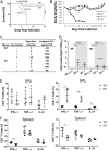
The NAGT mutant protects mice against a lethal homologous challenge. Vaccinated and unvaccinated (-) mice were challenged with a lethal dose of H1N1 (A/PR/8/34; 40 MLD50s) at 3 weeks postvaccination. (A) Survival rates were assessed daily for 14 days after challenge (log rank test). Each vaccinated group had 16 mice. (B) Weight loss in different mouse groups. (C) NP-specific antibody titers in mice at days 0, 7, and 14 after virus challenge. Antibody levels were studied by ELISA. (D) Lung virus titers in mice at days 3 and 7 postchallenge. Data from vaccinated mice are highlighted. DL, detection limit. (E and F) PR8-specific CD4+ and CD8+ T-cell recall responses in BAL fluid and spleens of infected mice at day 7 postinfection. Activities were determined by ICS assays. Data are means ± SD. *, P < 0.05; **, P < 0.01; ****, P < 0.0001.
FIG 6
FIG 6
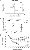
The NAGT mutant protects mice against a lethal heterosubtypic H3N2 challenge. Vaccinated and unvaccinated (-) mice were challenged with a lethal dose of H3N2 virus (HK68; 10 MLD50s) at 3 weeks postvaccination. (A) Survival rates were assessed daily for 14 days after challenge (log rank test). Each vaccinated group had ≥20 mice. (B) Weight loss in different mouse groups. (C) Lung virus titers in mice at days 3 and 7 postchallenge. Data from vaccinated mice are highlighted. DL, detection limit. (D) HK68-specific neutralizing antibody titers in mice before (3 weeks postvaccination) and after (days 7 and 14 postchallenge) challenge. Data from vaccinated mice are highlighted. (E) NP-specific antibody titers in mice at days 0, 7, and 14 after virus challenge. Antibody levels were studied by ELISA. (F and G) HK68-specific CD4+ and CD8+ T-cell recall responses in BAL fluid (F) and spleen tissue (G) of infected mice at day 7 postinfection. (H) Percentages of total DCs (left) and of CD11blo/− and CD11bhi DCs (right) in infected lung tissues at day 7 postchallenge. The dotted line shows the comparison between KO and WT mice at the same time point (t test). Data are means ± SD. *, P < 0.05; **, P < 0.01; ***, P < 0.001; ****, P < 0.0001.
FIG 7
FIG 7
The NAGT mutant protects mice against challenge with a lethal heterosubtypic highly pathogenic H5N1 virus. Vaccinated and unvaccinated (-) mice were challenged with a lethal dose of H5N1 virus (VN1203; 10 MLD50s) at 3 weeks postvaccination. (A) Survival rates were assessed daily for 14 days after challenge (log rank test). Each vaccinated group had 10 mice. (B) Lung virus titers in mice at day 7 postchallenge. DL, detection limit. (C) Weight loss in different mouse groups. Data are means ± SD. *, P < 0.05; **, P < 0.01; ****, P < 0.0001.
FIG 8
FIG 8
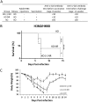
Enhanced protection by the NAGT mutant requires anti-α-Gal antibody in mice. (A) Anti-α-Gal antibody titers in mice immediately before vaccination and virus challenge. KO mice with (KO) or without (KO NR) rabbit RBC stimulation were vaccinated at 8 weeks of age. Mock-treated KO mice were used as controls [KO (-) NR]. Serum anti-α-Gal antibody titers in these mouse groups were determined by ELISA. (B). Survival rates of these mouse groups after a lethal HK68 virus challenge (10 MLD50s). Each group had 5 mice. (C) Weight loss in different mouse groups. Data are means ± SD. *, P < 0.05; ***, P < 0.001; ****, P < 0.0001.

References

    1. Krammer F, Smith GJD, Fouchier RAM, Peiris M, Kedzierska K, Doherty PC, Palese P, Shaw ML, Treanor J, Webster RG, Garcia-Sastre A. 2018. Influenza. Nat Rev Dis Primers 4:3. doi:10.1038/s41572-018-0002-y. - DOI - PMC - PubMed
    1. World Health Organization. 2009. Assessing the severity of an influenza pandemic. https://www.who.int/csr/disease/swineflu/assess/disease_swineflu_assess_....
    1. Rajao DS, Perez DR. 2018. Universal vaccines and vaccine platforms to protect against influenza Viruses in humans and agriculture. Front Microbiol 9:123. doi:10.3389/fmicb.2018.00123. - DOI - PMC - PubMed
    1. Paules CI, Sullivan SG, Subbarao K, Fauci AS. 2018. Chasing seasonal influenza—the need for a universal influenza vaccine. N Engl J Med 378:7–9. doi:10.1056/NEJMp1714916. - DOI - PubMed
    1. Boudreau CM, Alter G. 2019. Extra-neutralizing FcR-mediated antibody functions for a universal influenza vaccine. Front Immunol 10:440. doi:10.3389/fimmu.2019.00440. - DOI - PMC - PubMed

Publication types