. 2020 Mar 3;10(1):3930.
doi: 10.1038/s41598-020-60879-6.

Functional Consequences of PDK4 Deficiency in Doberman Pinscher Fibroblasts

Affiliations

Functional Consequences of PDK4 Deficiency in Doberman Pinscher Fibroblasts

Luiz Bolfer et al. Sci Rep. .

Abstract

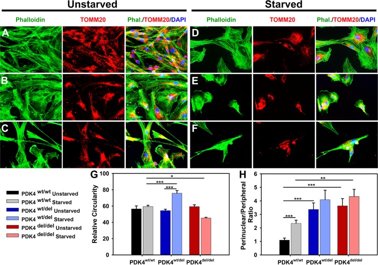
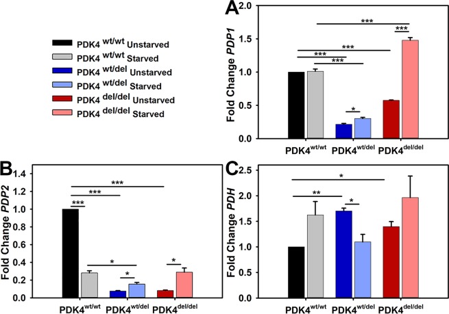
A splice site mutation in the canine pyruvate dehydrogenase kinase 4 (PDK4) gene has been shown to be associated with the development of dilated cardiomyopathy (DCM) in Doberman Pinchers (DPs). Subsequent studies have successfully demonstrated the use of dermal fibroblasts isolated from DPs as models for PDK4 deficiency and have shown activation of the intrinsic (mitochondrial mediated) apoptosis pathway in these cells under starvation conditions. For this study, we sought to further explore the functional consequences of PDK4 deficiency in DP fibroblasts representing PDK4wt/wt, PDK4wt/del, and PDK4del/del genotypes. Our results show that starvation conditions cause increased perinuclear localization of mitochondria and decreased cell proliferation, altered expression levels of pyruvate dehydrogenase phosphatase (PDP) and pyruvate dehydrogenase (PDH), dramatically increased PDH activity, and an impaired response to mitochondrial stress in affected cells. In sum, these results show the broad impact of PDK4 deficiency and reveal mechanistic pathways used by these cells in an attempt to compensate for the condition. Our data help to elucidate the mechanisms at play in this extremely prevalent DP disorder and provide further support demonstrating the general importance of metabolic flexibility in cell health.


Conflict of interest statement

The authors declare no competing interests.

Figures

Figure 1
Figure 1
DP fibroblast cellular morphology and mitochondrial localization. (A–C) Primary dermal fibroblasts from healthy controls PDK4wt/wt, heterozygous PDK4wt/del, or homozygous PDK4del/del, DPs were evaluated with IF staining of phalloidin (green) to reveal overall cellular architecture and the mitochondrial outer membrane protein TOMM20 (red) to locate mitochondria within cells. (D–F) Cells representing the three different genotypes were exposed to 24 hours of starvation conditions. (G) A graph indicating how relative circularity changed in both PDK4wt/del (increased) and PDK4del/del (decreased) cells in response to starvation conditions as compared to healthy controls under the same conditions. (H) Perinuclear localization of mitochondria was increased in both PDK4wt/del and PDK4del/del cells as compared to controls under the same condition except there was no significant difference between starved PDK4wt/del and PDK4wt/wt cells. (Data presented as mean + std. err. *p < 0.05, **p < 0.01, ***p < 0.001).
Figure 2
Figure 2
PDK isoform transcription levels in response to starvation. Relative transcription levels of (A) PDK1, (B) PDK2, (C) PDK3, and (D) PDK4 in cells representing each of the three groups PDK4wt/wt, PDK4wt/del, and PDK4del/del cells in unstarved and starved conditions. (Data presented as mean + std. err. *p < 0.05, **p < 0.01, ***p < 0.001).
Figure 3
Figure 3
PDP isoforms 1 and 2 and PDH transcription levels in response to starvation. Relative transcription levels of (A) PDP1, (B) PDP2, and (C) PDH, in cells representing each of the three groups PDK4wt/wt, PDK4wt/del, and PDK4del/del cells in unstarved and starved conditions. (Data presented as mean + std. err. *p < 0.05, **p < 0.01, ***p < 0.001).
Figure 4
Figure 4
PDH levels and phosphorylation status in response to starvation. (A) WB image using an antibody that recognizes all PDH and (B) quantified PDH data normalized to B-actin, and (C) WB image using an antibody that recognizes PDH in its phosphorylated state P-PDH and (D) corresponding quantified data normalized to B-actin. Quantified data represent 3 WBs of cell lysates representing each of the three groups PDK4wt/wt, PDK4wt/del, and PDK4del/del cells in unstarved and starved conditions. (E) Bar graph showing the ratio of P-PDH to PDH for each sample and condition. Full WB images are provided in supplemental data. (Data presented as mean + std. err. *p < 0.05).
Figure 5
Figure 5
Relative PDK4 expression and PDH activities. (A) Relative PDK4 expression levels as determined by an ELISA, (B) PDH activity levels between cells representing the three different genotypes and their responses to PDK4 and subsequently, glutamate additions (x-axis indicates minutes time). (C) Quantified PDH activity data showing significant differences between cells and conditions. PDK4wt/wt, PDK4wt/del, and PDK4del/del cells. (Data presented as mean + std. err. *p < 0.05, **p < 0.01, ***p < 0.001).
Figure 6
Figure 6
Extracellular Flux – Mitochondrial Stress Test. (A) Seahorse extracellular flux assay results profile showing relative responses to the addition of oligomycin, FCCP, and antimycin A/rotenone. (B) Quantified results representing relative OCR levels in PDK4wt/wt, PDK4wt/del, and PDK4del/del cells (Data presented as mean + std. err. *p < 0.05, **p < 0.01, ***p < 0.001).

References

    1. Hazebroek M, Dennert R, Heymans S. Idiopathic dilated cardiomyopathy: possible triggers and treatment strategies. Neth. Heart J. 2012;20:332–335. doi: 10.1007/s12471-012-0285-7. - DOI - PMC - PubMed
    1. Hazlett MJ, Maxie MG, Allen DG, Wilcock BP. A retrospective study of heart disease in doberman pinscher dogs. Can. Vet. J. 1983;24:205–210. - PMC - PubMed
    1. Lopes R, Solter PF, Sisson DD, Oyama MA, Prosek R. Characterization of canine mitochondrial protein expression in natural and induced forms of idiopathic dilated cardiomyopathy. Am. J. Vet. Res. 2006;67:963–970. doi: 10.2460/ajvr.67.6.963. - DOI - PubMed
    1. Lopes R, Solter PF, Sisson DD, Oyama MA, Prosek R. Correlation of mitochondrial protein expression in complexes I to V with natural and induced forms of canine idiopathic dilated cardiomyopathy. Am. J. Vet. Res. 2006;67:971–977. doi: 10.2460/ajvr.67.6.971. - DOI - PubMed
    1. Meurs KM, et al. A prospective genetic evaluation of familial dilated cardiomyopathy in the Doberman pinscher. J. Vet. Intern. Med. 2007;21:1016–1020. doi: 10.1111/j.1939-1676.2007.tb03058.x. - DOI - PubMed

Publication types

MeSH terms

Substances