Review
. 2020 May;16(5):276-283.
doi: 10.1038/s41574-020-0329-9. Epub 2020 Mar 3.

Insulin-PI3K signalling: an evolutionarily insulated metabolic driver of cancer

Affiliations
Review

Insulin-PI3K signalling: an evolutionarily insulated metabolic driver of cancer

Benjamin D Hopkins et al. Nat Rev Endocrinol. 2020 May.

Abstract

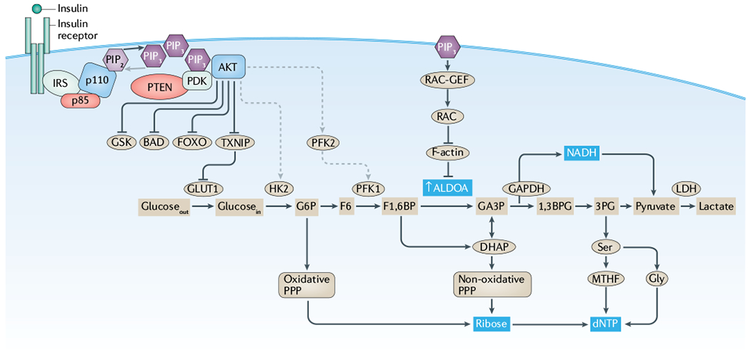
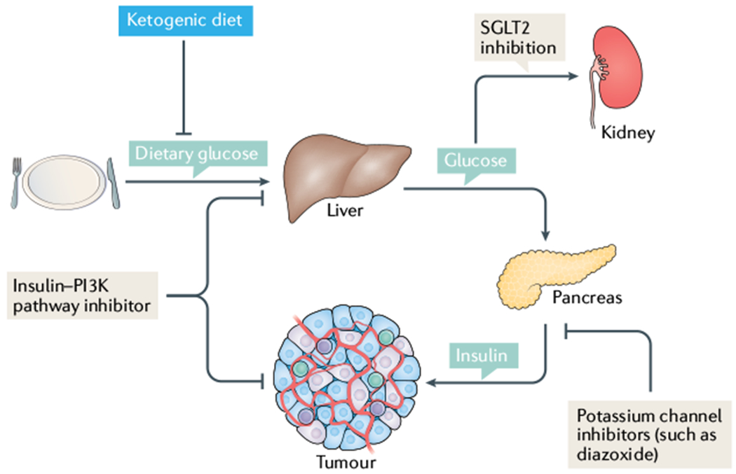
Cancer is driven by incremental changes that accumulate, eventually leading to oncogenic transformation. Although genetic alterations dominate the way cancer biologists think about oncogenesis, growing evidence suggests that systemic factors (for example, insulin, oestrogen and inflammatory cytokines) and their intracellular pathways activate oncogenic signals and contribute to targetable phenotypes. Systemic factors can have a critical role in both tumour initiation and therapeutic responses as increasingly targeted and personalized therapeutic regimens are used to treat patients with cancer. The endocrine system controls cell growth and metabolism by providing extracellular cues that integrate systemic nutrient status with cellular activities such as proliferation and survival via the production of metabolites and hormones such as insulin. When insulin binds to its receptor, it initiates a sequence of phosphorylation events that lead to activation of the catalytic activity of phosphoinositide 3-kinase (PI3K), a lipid kinase that coordinates the intake and utilization of glucose, and mTOR, a kinase downstream of PI3K that stimulates transcription and translation. When chronically activated, the PI3K pathway can drive malignant transformation. Here, we discuss the insulin-PI3K signalling cascade and emphasize its roles in normal cells (including coordinating cell metabolism and growth), highlighting the features of this network that make it ideal for co-option by cancer cells. Furthermore, we discuss how this signalling network can affect therapeutic responses and how novel metabolic-based strategies might enhance treatment efficacy for cancer.


Conflict of interest statement

Competing interests

B.D.H., M.D.G. and L.C.C. are all founders of and consultants for Faeth, a company developing nutrition for cancer care. L.C.C. is a founder and member of the scientific advisory board and board of directors of Agios and Petra Pharma, which are companies developing drugs to target metabolism.

Figures

Fig. 1 |
Fig. 1 |. Insulin regulates glycolysis through both AKT-dependent and AKT-independent mechanisms.
This simplified figure highlights components of the phosphoinositide 3-kinase (PI3K)-regulated cellular signalling events that occur downstream of the interaction between insulin and its receptor. While a myriad of growth factors and their cognate receptors (such as insulin growth factor or epidermal growth factor) activate PI3K signalling, here we highlight the insulin receptor, which is of particular importance to the systemic feedbacks central to this Review. The effects of insulin are mediated through both AKT-dependent (left side) and AKT-independent mechanisms (right side). From an evolutionary perspective, systemic insulin drives the intracellular PI3K signalling pathway to integrate systemic nutrient status and cellular metabolism with cellular functions, including proliferation and survival. This function enables insulin to coordinate cellular activities with systemic features, such as glucose availability, and provides a unified mechanism for the generation and utilization of key metabolites, such as deoxyribonucleotide triphosphates (dNTPs). Under normal conditions, this integration of systemic and cellular activity protects the organism by linking metabolic requirements and cellular growth, thereby constraining cellular activities based on systemic factors. In the context of oncogenic transformation, this integration makes the insulin–PI3K signalling axis a vulnerability, as activating mutations in oncogenes (encoding p110 and AKT) or inactivating mutations in tumour suppressors (encoding p85 and phosphatase and tensin homologue (PTEN)) can decouple this integration. IRS, insulin receptor substrate; LDH, lactate dehydrogenase; MTHF, 5,10-methenyltetrahydrofolate; PDK, phosphoinositide-dependent kinase; PIP2, phosphatidylinositol 4,5-bisphosphate; PIP3, phosphatidylinositol 3,4,5-trisphosphate; PPP, pentose phosphate pathway.
Fig. 2 |
Fig. 2 |. Tissue and tumour response to insulin signalling.
While canonical insulin signalling remains intact across tissue types, activation of the insulin receptor has tissue-specific effects. In muscle, insulin stimulates fatty acid oxidation as well as glucose uptake and utilization. By contrast, in adipose tissues and liver, insulin stimulates a storage phenotype, driving fatty acid production, inhibiting lipolysis in adipose tissue and inducing glucose uptake while suppressing gluconeogenesis in the liver. Similarly, tumours can use insulin signalling to drive glucose uptake as well as inducing cell survival and proliferation.
Fig. 3 |
Fig. 3 |. Analysis of the frequency of mutations in insulin–PI3K signalling.
Histogram of the frequency of mutations for the 32 The Cancer Genome Atlas (TCGA) pan-cancer data sets from cBioPortal, showing that the distribution of mutations in genes (PIK3CA, PIK3R1, AKT1, AKT2, AKT3, PTEN and INSR) encoding elements of the insulin–phosphoinositide 3-kinase (PI3K) cascade are not uniform across tumour types. These data highlight the frequency with which this pathway is altered in human cancer and support the idea that different tumour types might be differentially sensitive to genetic modulations of this pathway as the result of the tissues from which they arise or the specific environment of that tissue. Looking across the data set, which covers sequencing from 32 studies, demonstrates the high frequency with which components of this signalling cascade are altered, particularly p110α (encoded by PIK3CA, p110α is a catalytic subunit of PI3K) and phosphatase and tensin homologue (the phosphatase that catalyses the reverse reaction). In total, 38% of the cases in the TCGA pan-cancer studies had mutations in these seven genes.
Fig. 4 |
Fig. 4 |. Mechanisms to inhibit insulin feedback in tumours.
In a patient with cancer who is on a normal carbohydrate-replete diet, normal glucose homeostasis leads to glucose uptake and storage in the liver. When insulin signalling is not active, the liver induces gluconeogenesis to maintain systemic blood levels of glucose. In the clinic, when patients are given inhibitors that target the insulin receptor, phosphoinositide 3-kinase (PI3K) or AKT, their tissues perceive this as a decrease in insulin levels. In the liver, this means increasing the release of glucose, which in turn raises blood levels of glucose and induces increased insulin release from the pancreas. As insulin can act as a mitogen and regulate tumour cell survival signalling, this systemic feedback might be detrimental to treatment efficacy. By administering these agents in combination with sodium–glucose co-transporter 2 (SGLT2) inhibitors that cause the removal of glucose by the kidney, or a ketogenic diet that removes dietary carbohydrate so there are insufficient stores in the liver to cause notable rises in blood levels of glucose, one can prevent therapy-induced spikes in insulin levels, which might limit treatment efficacy. More broadly, limiting insulin exposure in patients might decrease survival and proliferative signalling in tumours where these features are typically induced by systemic insulin.

References

    1. Karamitsos DT The story of insulin discovery. Diabetes Res. Clin. Pract 93, S2–S8 (2011). - PubMed
    1. Banting FG & Best CH Pancreatic Extracts (Toronto Univ. Library, 1922).
    1. Pollak M Insulin and insulin-like growth factor signalling in neoplasia. Nat. Rev. Cancer 8, 915–928 (2008). - PubMed
    1. Dong MQ et al. Quantitative mass spectrometry identifies insulin signaling targets in C. elegans. Science 317, 660–663 (2007). - PubMed
    1. Engelman JA, Luo J & Cantley LC The evolution of phosphatidylinositol 3-kinases as regulators of growth and metabolism. Nat. Rev. Genet 7, 606–619 (2006). - PubMed

Publication types