

Actinin-4 splice variant - a complementary diagnostic and prognostic marker of pancreatic neuroendocrine neoplasms
- PMID: 32127958
- PMCID: PMC7052930
- DOI: 10.7150/jca.37503
Actinin-4 splice variant - a complementary diagnostic and prognostic marker of pancreatic neuroendocrine neoplasms
Abstract
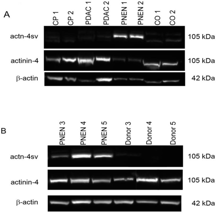
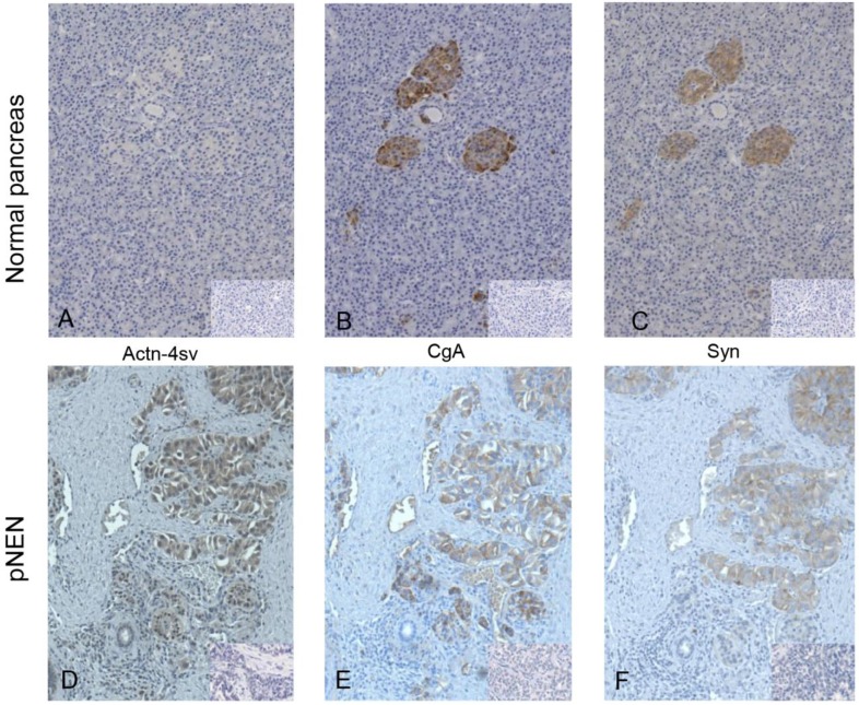
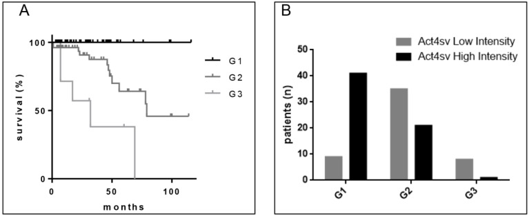
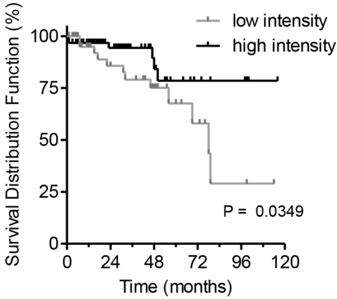
Introduction: For pathological diagnosis of pancreatic neuroendocrine neoplasms (pNENs) the routinely used immunohistochemical markers are chromogranin A (CgA) and synaptophysin (Syn). Their ability as prognostic markers is not well established. A splice variant of actinin-4 (Actn-4sv) was recently found to be an excellent biomarker of neuroendocrine neoplasms of the lung. We aimed to investigate the expression of Actn-4sv in pNENs and evaluate its quality as a biomarker of pNENs. Methods: Paraffin-embedded and frozen tissues specimens from 122 pNENs were analyzed. Western blots were performed to prove and compare the relative amount of Actn-4sv expression in pNENs tissue homogenates. For comparison pancreatic ductal adenocarcinoma (PDAC) and normal pancreatic tissues were analyzed in parallel. Immunohistochemistry (IHC) of paraffin sections of pNENs for Actn-4sv were performed and compared to the classic neuroendocrine markers CgA and Syn. Correlations were calculated between the staining intensity and distribution of Actn-4sv and staging, grading and afflicted lymph nodes respectively. Results: Actn-4sv was expressed in 88.5% (108/122) of pNENs, but not in normal pancreatic tissues (0/14) or PDAC (0/14). Compared to CgA and Syn, Actn-4sv was not detectable in islet cells of the normal pancreas. Staining intensity of Actn-4sv on pNENs negatively correlated to the histological grading (Spearman r=-0.4990, p<0.0001) and staging (r = -0.2581, p = 0.0041) but no correlation to afflicted lymph nodes was found. A significantly better overall survival was observed for pNEN patients with higher expression of Actn-4sv (hazard ratio 2.7; log-rank test p= 0.0349). Conclusions: The expression of Actn-4sv may be an important prognostic factor for patients with pNENs. Its expression correlates with the grading and staging of the tumors.
Keywords: actinin-4; actinin-4 splice variant; pNEN; survival.
© The author(s).
Conflict of interest statement
Competing Interests: The authors have declared that no competing interest exists.
Figures
Similar articles
-
Comparison of morphological features in lymph node metastasis between pancreatic neuroendocrine neoplasms and pancreatic ductal adenocarcinomas.Pancreatology. 2020 Jul;20(5):936-943. doi: 10.1016/j.pan.2020.05.013. Epub 2020 May 22. Pancreatology. 2020. PMID: 32553561
-
CD56 Expression Is Associated with Biological Behavior of Pancreatic Neuroendocrine Neoplasms.Cancer Manag Res. 2020 Jun 17;12:4625-4631. doi: 10.2147/CMAR.S250071. eCollection 2020. Cancer Manag Res. 2020. PMID: 32606955 Free PMC article.
-
The added value of intravoxel incoherent motion diffusion weighted imaging parameters in differentiating high-grade pancreatic neuroendocrine neoplasms from pancreatic ductal adenocarcinoma.Oncol Lett. 2019 Nov;18(5):5448-5458. doi: 10.3892/ol.2019.10863. Epub 2019 Sep 13. Oncol Lett. 2019. PMID: 31612053 Free PMC article.
-
[Surgical strategies for small sporadic neuroendocrine pancreatic tumors].Chirurg. 2018 Jun;89(6):422-427. doi: 10.1007/s00104-018-0632-3. Chirurg. 2018. PMID: 29637243 Review. German.
-
Update on surgical treatment of pancreatic neuroendocrine neoplasms.World J Gastroenterol. 2014 Oct 14;20(38):13893-8. doi: 10.3748/wjg.v20.i38.13893. World J Gastroenterol. 2014. PMID: 25320524 Free PMC article. Review.
Cited by
-
Altered splicing machinery in lung carcinoids unveils NOVA1, PRPF8 and SRSF10 as novel candidates to understand tumor biology and expand biomarker discovery.J Transl Med. 2023 Dec 4;21(1):879. doi: 10.1186/s12967-023-04754-8. J Transl Med. 2023. PMID: 38049848 Free PMC article.
-
The uprise of RNA biology in neuroendocrine neoplasms: altered splicing and RNA species unveil translational opportunities.Rev Endocr Metab Disord. 2023 Apr;24(2):267-282. doi: 10.1007/s11154-022-09771-4. Epub 2022 Nov 24. Rev Endocr Metab Disord. 2023. PMID: 36418657 Free PMC article. Review.
References
-
- Milan SA, Yeo CJ. Neuroendocrine tumors of the pancreas. Curr Opin Oncol. 2012;24:46–55. - PubMed
-
- Schimmack S, Svejda B, Lawrence B, Kidd M, Modlin IM. The diversity and commonalities of gastroenteropancreatic neuroendocrine tumors. Langenbeck's archives of surgery. 2011;396:273–98. - PubMed
-
- Lawrence B, Gustafsson BI, Chan A, Svejda B, Kidd M, Modlin IM. The epidemiology of gastroenteropancreatic neuroendocrine tumors. Endocrinology and metabolism clinics of North America. 2011;40:1–18. - PubMed
-
- Kwekkeboom DJ, Krenning EP, Scheidhauer K, Lewington V, Lebtahi R, Grossman A. et al. ENETS Consensus Guidelines for the Standards of Care in Neuroendocrine Tumors: somatostatin receptor imaging with (111)In-pentetreotide. Neuroendocrinology. 2009;90:184–9. - PubMed
-
- Sundin A, Vullierme MP, Kaltsas G, Plockinger U, Mallorca Consensus Conference p, European Neuroendocrine Tumor S. ENETS Consensus Guidelines for the Standards of Care in Neuroendocrine Tumors: radiological examinations. Neuroendocrinology. 2009;90:167–83. - PubMed
LinkOut - more resources
Full Text Sources
Research Materials
Miscellaneous
