

MIFlowCyt-EV: a framework for standardized reporting of extracellular vesicle flow cytometry experiments
- PMID: 32128070
- PMCID: PMC7034442
- DOI: 10.1080/20013078.2020.1713526
MIFlowCyt-EV: a framework for standardized reporting of extracellular vesicle flow cytometry experiments
Abstract
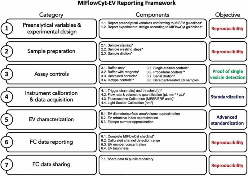
Extracellular vesicles (EVs) are small, heterogeneous and difficult to measure. Flow cytometry (FC) is a key technology for the measurement of individual particles, but its application to the analysis of EVs and other submicron particles has presented many challenges and has produced a number of controversial results, in part due to limitations of instrument detection, lack of robust methods and ambiguities in how data should be interpreted. These complications are exacerbated by the field's lack of a robust reporting framework, and many EV-FC manuscripts include incomplete descriptions of methods and results, contain artefacts stemming from an insufficient instrument sensitivity and inappropriate experimental design and lack appropriate calibration and standardization. To address these issues, a working group (WG) of EV-FC researchers from ISEV, ISAC and ISTH, worked together as an EV-FC WG and developed a consensus framework for the minimum information that should be provided regarding EV-FC. This framework incorporates the existing Minimum Information for Studies of EVs (MISEV) guidelines and Minimum Information about a FC experiment (MIFlowCyt) standard in an EV-FC-specific reporting framework (MIFlowCyt-EV) that supports reporting of critical information related to sample staining, EV detection and measurement and experimental design in manuscripts that report EV-FC data. MIFlowCyt-EV provides a structure for sharing EV-FC results, but it does not prescribe specific protocols, as there will continue to be rapid evolution of instruments and methods for the foreseeable future. MIFlowCyt-EV accommodates this evolution, while providing information needed to evaluate and compare different approaches. Because MIFlowCyt-EV will ensure consistency in the manner of reporting of EV-FC studies, over time we expect that adoption of MIFlowCyt-EV as a standard for reporting EV- FC studies will improve the ability to quantitatively compare results from different laboratories and to support the development of new instruments and assays for improved measurement of EVs.
Keywords: Extracellular vesicles; flow cytometry; framework; reporting; standardization.
© 2020 The Author(s). Published by Informa UK Limited, trading as Taylor & Francis Group on behalf of The International Society for Extracellular Vesicles.
Figures
References
-
- Fais S, O’Driscoll L, Borras FE, et al. Evidence-based clinical use of nanoscale extracellular vesicles in nanomedicine. ACS Nano. 2016;10(4):3886–17. - PubMed
Grants and funding
LinkOut - more resources
Full Text Sources
Other Literature Sources
