. 2020 Feb 14;5(1):15.
doi: 10.1038/s41541-020-0164-y. eCollection 2020.

A single dose polyanhydride-based nanovaccine against paratuberculosis infection

Affiliations

A single dose polyanhydride-based nanovaccine against paratuberculosis infection

Akanksha Thukral et al. NPJ Vaccines. .

Abstract

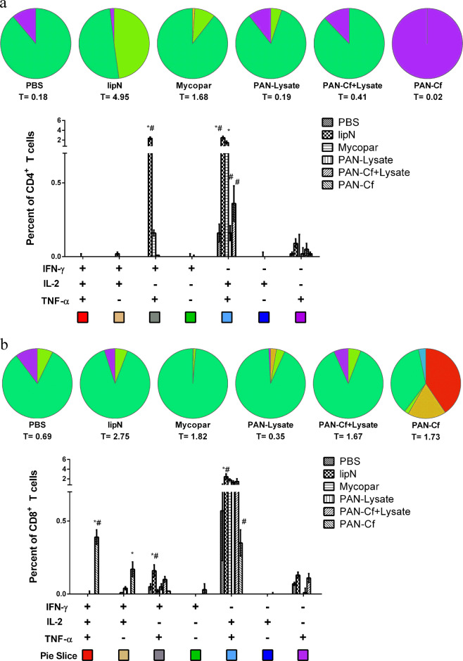
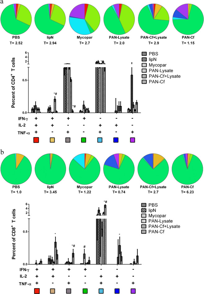
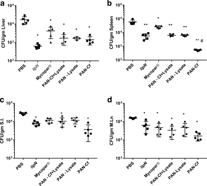
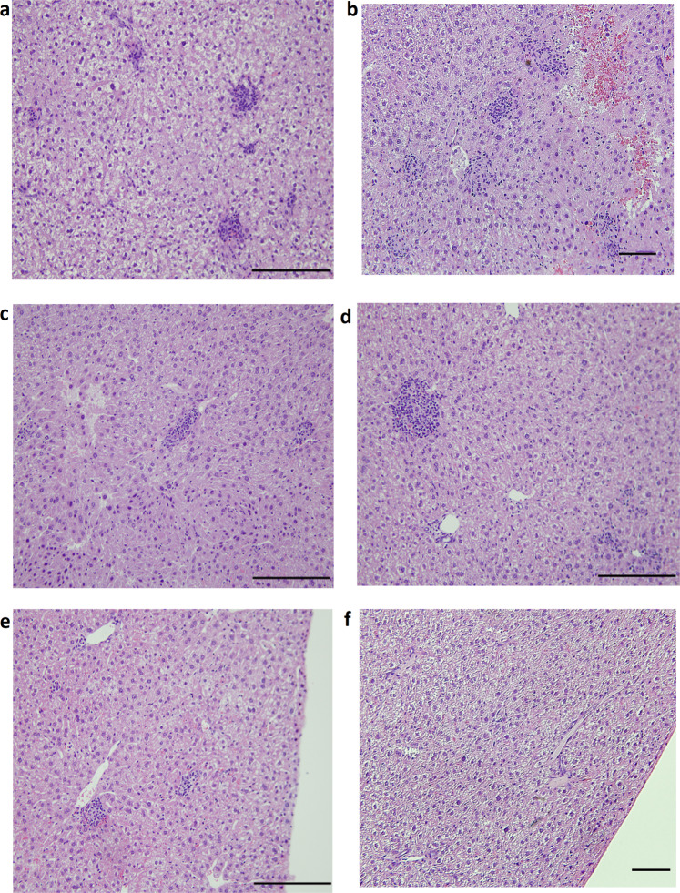
Mycobacterium avium subsp. paratuberculosis (M. paratuberculosis) causes Johne's disease in ruminants and is characterized by chronic gastroenteritis leading to heavy economic losses to the dairy industry worldwide. The currently available vaccine (inactivated bacterin in oil base) is not effective in preventing pathogen shedding and is rarely used to control Johne's disease in dairy herds. To develop a better vaccine that can prevent the spread of Johne's disease, we utilized polyanhydride nanoparticles (PAN) to encapsulate mycobacterial antigens composed of whole cell lysate (PAN-Lysate) and culture filtrate (PAN-Cf) of M. paratuberculosis. These nanoparticle-based vaccines (i.e., nanovaccines) were well tolerated in mice causing no inflammatory lesions at the site of injection. Immunological assays demonstrated a substantial increase in the levels of antigen-specific T cell responses post-vaccination in the PAN-Cf vaccinated group as indicated by high percentages of triple cytokine (IFN-γ, IL-2, TNF-α) producing CD8+ T cells. Following challenge, animals vaccinated with PAN-Cf continued to produce significant levels of double (IFN-γ, TNF-α) and single cytokine (IFN-γ) secreting CD8+ T cells compared with animals vaccinated with an inactivated vaccine. A significant reduction in bacterial load was observed in multiple organs of animals vaccinated with PAN-Cf, which is a clear indication of protection. Overall, the use of polyanhydride nanovaccines resulted in development of protective and sustained immunity against Johne's disease, an approach that could be applied to counter other intracellular pathogens.

Keywords: Adjuvants; Immunology; Vaccines.


Conflict of interest statement

Competing interestsDr. Adel M. Talaat has an ownership interest in Pan Genome Systems, INC, which is working in the area of animal vaccine development. Also, Dr. Yashdeep Phanse is currently employed by the same company.

Figures

Fig. 1
Fig. 1. Experimental design for vaccination and challenge.
Five- to eight-week-old female C57BL/6 mice were vaccinated with subcutaneous injection and challenged six weeks later with virulent strain of M. paratuberculosis JTC-1285 by the intraperitoneal route. Mice (n = 5/group) were sacrificed at various time points. Tissues and blood samples were collected to measure bacterial burden, cytokine levels and histopathology.
Fig. 2
Fig. 2. Characterization of M. paratuberculosis antigen-containing nanoparticle-based vaccines.
Scanning electron microscopy images of 20:80 CPTEG:CPH nanoparticles loaded with 2.5% whole cell lysate (a) and 2.5% culture filtrate (b) showed similar spherical morphology and size (~200 nm) as blank nanoparticles (c). Scale bar = 1 μm.
Fig. 3
Fig. 3. Pre-challenge immune response specific to lysate of M. paratuberculosis.
C57BL/6 mice (n = 5) were immunized with various vaccine groups and 6 WPV, five mice from each group were euthanized. Spleens were harvested; lymphocytes were isolated and stimulated with the M. paratuberculosis lysate for 24 h. Cells were then stained for CD4+ (a) and CD8+ (b) cell surface markers and intracellular cytokines. The total percentage of T cells secreting particular cytokines are indicated below each pie chart (denoted by T = number). The error bars show the standard error of the mean for five individually analyzed mice. * indicates p < 0.05; ** indicates p < 0.001. * denotes comparison with PBS while # denotes comparison with Mycopar®. Results were expressed as the increase in the percentage of the cells with positive staining relative to that of an unstimulated sample stained with the same antibody.
Fig. 4
Fig. 4. Early cellular responses in vaccine groups following challenge with a wild type strain of M. paratuberculosis.
Six to eight week-old C57BL/6 mice were immunized with various vaccine candidates. At 6 WPV they were challenged with M. paratuberculosis JTC-1285 and euthanized 12 weeks later (12 WPC). The lymphocytes were isolated from the spleens and stimulated with whole cell lysate of M. paratuberculosis for 24 h. Cells were then stained for CD4+ (a) and CD8+ (b) cell surface markers and intracellular cytokines and were measured by flow cytometry. The total percentage of T cells secreting particular cytokines are indicated below each pie chart (denoted by T = number). The error bars show the standard error of the mean for five individually analyzed mice. * indicates p < 0.05; ** indicates p < 0.001. * denotes comparison with PBS while # denotes comparison with Mycopar®. Results were expressed as the increase in the percentage of the cells with positive staining relative to that of an unstimulated sample stained with the same antibody.
Fig. 5
Fig. 5. Protection against challenge strain of M. paratuberculosis.
Levels of M. paratuberculosis colonization in body organs of mice at 12 WPC. The total colony counts for each individual animal for each vaccine groups are shown in spleen (a), liver (b), small intestine (c) and mesenteric lymph node (d). Error bars indicate standard deviation. * indicates p < 0.05; ** indicates p < 0.001. * denotes comparison with PBS while # denotes comparison with Mycopar®.
Fig. 6
Fig. 6. Histopathology of vaccinated and challenged mice.
Liver tissues from PBS (a), Mycopar® (b), lipN (c), PAN-Lysate (d), PAN-Cf + Lysate (e), PAN-Cf (f) vaccinated mice were harvested at 12 WPC. Tissues were sectioned to 5 µm slices and stained with H&E and analyzed at 20× magnification. Scale bar: 100 µm.

References

    1. Hendrick SH, et al. Effect of paratuberculosis on culling, milk production, and milk quality in dairy herds. J. Am. Vet. Med. Assoc. 2005;227:1302–1308. - PubMed
    1. Lombard J, et al. Herd-level prevalence of Mycobacterium avium subsp. paratuberculosis infection in United States dairy herds in 2007. Preventive Vet. Med. 2013;108:234–238. - PubMed
    1. Raizman EA, Fetrow JP, Wells SJ. Loss of income from cows shedding Mycobacterium avium subspecies paratuberculosis prior to calving compared with cows not shedding the organism on two Minnesota dairy farms. J. Dairy Sci. 2009;92:4929–4936. - PubMed
    1. Ott SL, Wells SJ, Wagner BA. Herd-level economic losses associated with Johne’s disease on US dairy operations. Prev. Vet. Med. 1999;40:179–192. - PubMed
    1. Hasonova L, Pavlik I. Economic impact of paratuberculosis in dairy cattle herds: a review. Veterinarni Med. 2006;51:193–211.

LinkOut - more resources