. 2020 Feb 14;5(1):16.
doi: 10.1038/s41541-020-0163-z. eCollection 2020.

Modified mRNA/lipid nanoparticle-based vaccines expressing respiratory syncytial virus F protein variants are immunogenic and protective in rodent models of RSV infection

Affiliations

Modified mRNA/lipid nanoparticle-based vaccines expressing respiratory syncytial virus F protein variants are immunogenic and protective in rodent models of RSV infection

Amy S Espeseth et al. NPJ Vaccines. .

Abstract

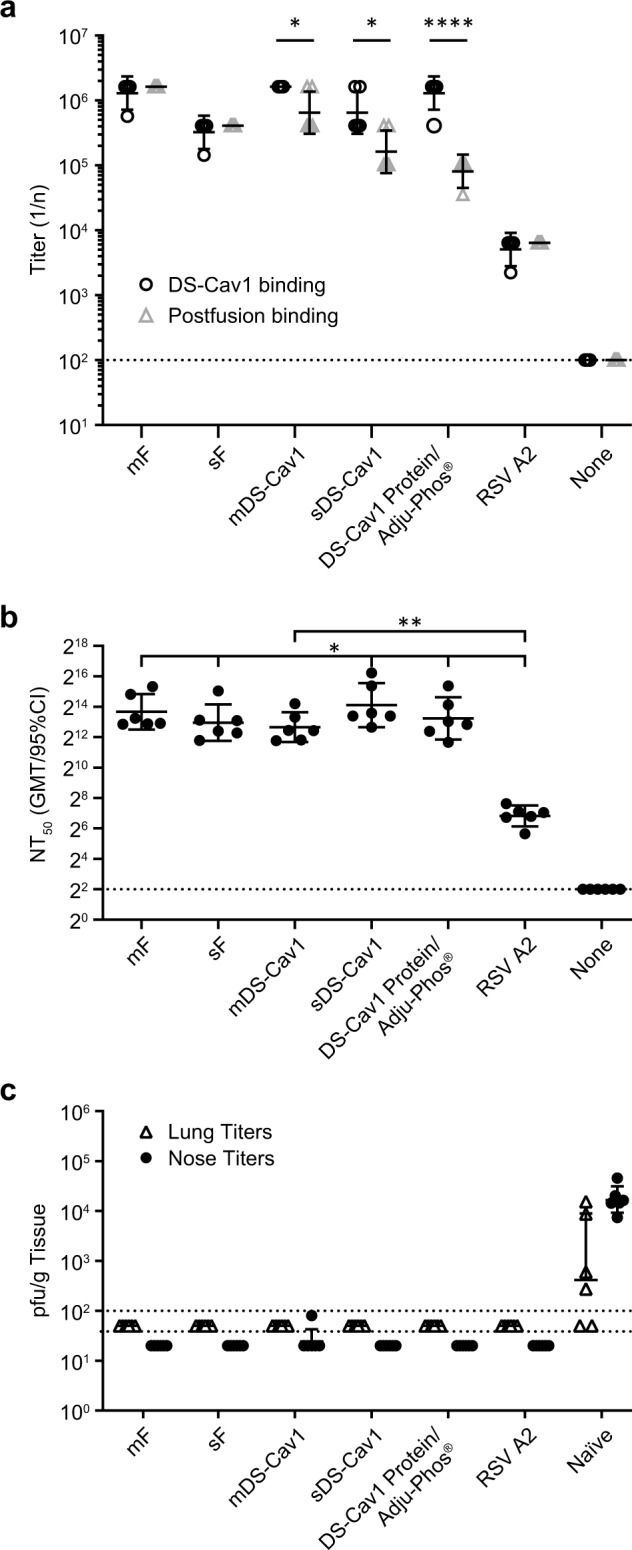
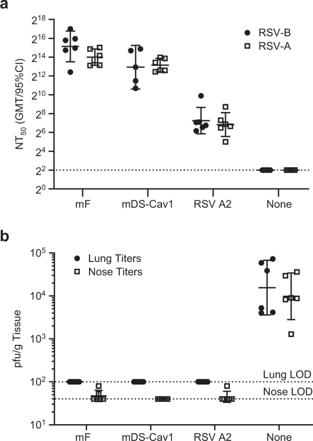
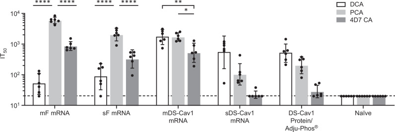
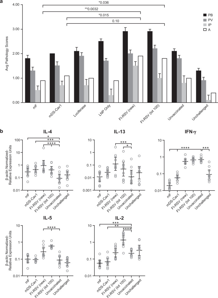
The RSV Fusion (F) protein is a target for neutralizing antibody responses and is a focus for vaccine discovery; however, the process of RSV entry requires F to adopt a metastable prefusion form and transition to a more stable postfusion form, which displays less potent neutralizing epitopes. mRNA vaccines encode antigens that are translated by host cells following vaccination, which may allow conformational transitions similar to those observed during natural infection to occur. Here we evaluate a panel of chemically modified mRNA vaccines expressing different forms of the RSV F protein, including secreted, membrane associated, prefusion-stabilized, and non-stabilized structures, for conformation, immunogenicity, protection, and safety in rodent models. Vaccination with mRNA encoding native RSV F elicited antibody responses to both prefusion- and postfusion-specific epitopes, suggesting that this antigen may adopt both conformations in vivo. Incorporating prefusion stabilizing mutations further shifts the immune response toward prefusion-specific epitopes, but does not impact neutralizing antibody titer. mRNA vaccine candidates expressing either prefusion stabilized or native forms of RSV F protein elicit robust neutralizing antibody responses in both mice and cotton rats, similar to levels observed with a comparable dose of adjuvanted prefusion stabilized RSV F protein. In contrast to the protein subunit vaccine, mRNA-based vaccines elicited robust CD4+ and CD8+ T-cell responses in mice, highlighting a potential advantage of the technology for vaccines requiring a cellular immune response for efficacy.

Keywords: RNA vaccines; Vaccines; Virology.


Conflict of interest statement

Competing interestsA.S.E., P.J.C., M.P.C., D.W., D.J.D., C.C., G.O., J.G., R.S., S.T., Z.W., J.A., L.Z., J.A.F., K.S.C., D.C.F., K.V., A.H.L., J.S.S., M.E.G., D.H., A.J.B. are all employees and/or stockholders of Merck & Co., Inc., Kenilworth, NJ, USA. C.A.S., K.B., and G.C. are employees and/or stockholders of Moderna Inc., Cambridge, MA, USA. G.C., K.B., A.S.E., D.W. and A.J.B. are co-inventors on a patent application related to this work.40

Figures

Fig. 1
Fig. 1. Variants of RSV F protein expressed by mRNA vaccines.
The different forms of RSV protein expressed by the modified mRNA vaccines evaluated in this study are shown. wtRSV (mF) expresses the full length RSV F protein from the RSV A2 strain. The locations of the signal peptide (SP), p27 peptide (p27), fusion peptide (FP), heptad repeat A (HRA), heptad repeat B (HRB), and transmembrane peptide (TM) are shown. For each construct, arrows indicated proteolytic cleavage sites that remain in the expressed protein. Deleted amino acid sequences are indicated with a horizontal line replacing the bar. Point mutations added to stabilize the prefusion conformation of the antigen are indicated with a solid circle (•) and the identity of the amino acid change is indicated below the circle. The newly generated disulfide bonds formed in the DS-Cav1-based constructs is indicated with a bracket. Secreted constructs in which the transmembrane domain and cytoplasmic sequence are deleted and replaced with a foldon or GCN4 peptide sequence to support trimerization are indicated with a gray or black-filled bar, respectively.
Fig. 2
Fig. 2. mRNA/LNP vaccines elicit conformation-indicating antibody titers in mouse.
BALB/c mice (N = 5–6) were immunized twice with 10 μg mRNA/LNP vaccine or DS-Cav1 protein formulated with Adju-Phos®. Sera taken 2 weeks following the second immunization was assayed for the presence of D25-competing antibodies (DCA, (gray bar), palivizumab-competing antibodies (PCA, (striped bar), or 4D7 competing antibodies (4D7 CA, (white bar)). The geometric mean antibody titer needed to inhibit 50% of mAb (D25, palivizumab, or 4D7) binding to RSV F protein (IT50) is shown, along with the 95% confidence interval (CI). The dotted line indicates the limit of detection of the competition ELISA. DCA, PCA and 4D7 competing titers were compared within each group by one-way ANOVA using Graph Pad Prism software. Differences between titers within each immunization group are shown (p < 0.05 = *, p < 0.01 = **).
Fig. 3
Fig. 3. mRNA/LNP vaccines expressing RSV F elicit cellular immune responses in BALB/c mice.
BALB/c mice (N = 5–6) were immunized twice with 10 μg mRNA/LNP vaccine or DS-Cav1 protein formulated with Adju-Phos®. Spleens were harvested 4 weeks following the second immunization. Splenocytes were incubated with pooled RSV F peptides and anti-mouse CD28 and were then stained with antibodies to the cell surface markers CD3, CD4, and CD8, as well as with a panel of anticytokine antibodies. The fluorescently tagged cells were then analyzed by flow cytometry using the gating strategy summarized in Supplementary Fig. 3. The percent of CD4+ T-cells (a) and CD8+ T-cells (b) responding to RSV F peptides with production of IFN-γ (white bar), IL-2 (light gray bar), or TNF-α (dark gray bar) is shown. Measurements for each animal are indicated (•). The height of the bar in the graph indicates the geometric mean calculation for the group ± 95% CI. *p < 0.05, **p < 0.01, ***p < 0.001, ****p < 0001 compared with the DS-Cav1 protein group by two-sided unpaired T-test with Welch’s correction. The dark gray dotted line indicates the limit of detection of the assay.
Fig. 4
Fig. 4. mRNA/LNP vaccines expressing RSV F are immunogenic and protective against RSV-A challenge in cotton rats.
Cotton rats (N = 6) were immunized twice with 25 μg mRNA/LNP vaccine or DS-Cav1 formulated with Adju-Phos®. a Sera collected 2 weeks following the second immunization were assayed for binding to the prefusion DS-Cav1 protein (white circle) or postfusion F protein (white triangle) by ELISA. Endpoint titers are shown for each animal and the center bar indicates the geometric mean and error bars show the 95% confidence interval for each group. The dotted line indicates the limit of detection for the assay. Binding titers to prefusion versus postfusion F protein were compared for each group by two-sided unpaired t-test using GraphPad Prism software. Cotton rats immunized with mDS-Cav1, sDS-Cav1, or DS-Cav1 protein had significantly higher binding to the DS-Cav1 prefusion F protein (p = 0.01, 0.04, <0.0001, respectively). b Sera from each animal was assayed for the ability to neutralize RSV Long infection in vitro. The 50% neutralizing titer (NT50) was defined as the reciprocal of the serum dilution required to neutralize 50% of input virus as determined by four-parameter sigmoidal curve fit. Measurements for each animal are indicated (black circle). The center bar indicates the geometric mean titers and error bars show the 95% confidence interval. The limit of detection titer of 4 is indicated by a dashed line. Neutralizing titers for each immunized group were compared to the control group given RSV A2 intranasally by two-sided unpaired t-test using GraphPad Prism software and significant differences are shown on the graph. Immunization with mRNA vaccines or DS-Cav1 protein led to significantly higher neutralizing antibody titers than intranasal RSV A2 (sDS-Cav1, p = 0.04; mDS-Cav1 p = 0.0093; sF, p = 0.04; mF, p = 0.02; DS-Cav1 protein, p = 0.04). No other significant differences between vaccinated groups were identified. c Animals were challenged with RSV A2 4 weeks after the second immunization. Nose and lung tissue were collected 4 days following challenge and assayed for the presence of RSV by plaque assay. Lung titers (∆) and nose titers (•) are shown for each animal. Geometric mean titer with 95% CI is shown for each group. The assay limit of detection (LOD) is 100 for lung titers and 40 for nose titers; each is indicated on the graph with a dotted line. Animals with no RSV infection measured were plotted at one-half the LOD. Because the viral titers in the lungs of the unvaccinated, RSV-challenged animals were lower than expected, qPCR to measure RSV RNA levels was conducted on the animals in this study. The qPCR data confirmed that the animals received an effective RSV challenge and the mRNA vaccines reduced RSV titer in the lung and nose, comparable to the RSV A2 group (Supplementary Fig. 4).
Fig. 5
Fig. 5. mRNA vaccines expressing RSV mF and mDS-Cav1 neutralize both RSV A and RSV B and are protective against RSV B challenge in cotton rats.
Cotton rats (N = 6) were immunized twice with 25 μg mRNA/LNP vaccine or DS-Cav1 formulated with Adju-Phos®. Sera was isolated 4 weeks following the second immunization and assayed for neutralization of RSV A (Long) and RSV B (18537). a NT50 titers calculated as described above for RSV-A (Long) (white square) and RSV-B (18537) (black circle) are plotted for each group. The center bar indicates the geometric mean titers and error bars show the 95% confidence interval. The limit of detection titer of 4 is indicated by a dashed line. No difference between NT50 titers for RSV-A and RSV-B was detected for any of the groups by two-sided paired t-test. b Animals were challenged with RSV B 18537 4 weeks after the second immunization. Nose and lung tissue were collected 4 days following challenge and assayed for the presence of RSV by plaque assay. Lung titers (black circle) and nose titers (white square) are shown for each animal. Geometric mean titer with 95% CI is shown for each group. The assay limit of detection (LOD) is 100 for lung titers and 40 for nose titers; each is indicated on the graph with a dotted line. Animals with no RSV infection measured were plotted at the LOD.
Fig. 6
Fig. 6. mRNA vaccines expressing prefusion RSV F elicit higher DCA titers than unmodified full length or secreted forms of RSV F in cotton rats.
Cotton rats (N = 6) were immunized twice with 25 μg mRNA/LNP vaccine or DS-Cav1 formulated with Adju-Phos®. Sera collected 2 weeks following the second immunization were assayed for DCA (white bar), PCA (light gray bar), or 4D7CA (dark gray bar). The IT50 for each animal is plotted, and the geometric mean and 95% confidence intervals is shown. The dotted line at IT50 = 20 indicates the LOD for the assay. Animals with no competing antibody titers were assigned a titer at the LOD. DCA, PCA and 4D7 competing titers were compared within each group by one-way ANOVA using Graph Pad Prism software. Differences between titers within each immunization group are shown (p < 0.05 = *, p < 0.01 = **, p < 0.001 = ***, p < 0.0001 = ****).
Fig. 7
Fig. 7. RSV F-mRNA Vaccines do not lead to VERD in cotton rat.
Cotton rats (N = 10) were immunized twice with 25 μg mF mRNA/LNP, mDS-Cav1 mRNA/LNP, Luciferase mRNA/LNP, LNP only (no RNA), FI-RSV (lot 100), FI-RSV generated using similar methods to lot 100 (FI-RSV (new)) or were left unvaccinated. With the exception of one unvaccinated group, all groups were challenged 4 weeks following the second immunization with RSV A2. Five days following the challenge, the animals were sacrificed. Lungs from each animal were trisected and processed for virus quantification (Supplementary Fig. 5b, c), pathology (a), and cytokine mRNA analysis (b). a Lung pathology following RSV challenge. Lung tissue from each animal was embedded in paraffin, sectioned, stained with hematoxylin and eosin, and given a pathology score from 0 (no pathology) to 4 (severe pathology) by a blinded pathologist. Sections were scored for peribronchiolitis (PB), perivasculitis (PV), interstitial pneumonia (IP), and alveolitis (A). Mean pathology score with standard error is shown. Raw histopathology scores for each animal are shown in Supplementary Table 1. Lung pathology scores were assessed using a two-sided exact permutation test. The mF and mDS-Cav1 mRNA vaccines resulted in significantly lower levels of pathology relative to both FI-RSV controls (for alveolitis, p = 0.0032 for mF versus FI-RSV (new) and 0.0358 for mF versus FI-RSV lot 100; p = 0.0151 for mDS-Cav1 versus FI-RSV (new) and 0.1016 for mDS-Cav1 versus FI-RSV lot 100), and the pathology scores for these two groups are not significantly different from the luciferase mRNA/LNP, LNP alone, unvaccinated and unchallenged negative control groups. b Cytokine gene expression after vaccination and RSV challenge. mRNA was isolated from the lungs of each cotton rat and IL-4, IL-13, IL-5, IFN-γ, and IL-2 mRNA levels were determined by rtPCR. The relative level of each mRNA was determined by normalizing to levels of the housekeeping gene β-actin and plotted for each animal. The geometric mean and 95% confidence interval for the β-actin normalized relative mRNA expression units for each of the five cytokines is shown. Differences between the groups were determined by two-sided t-test using the error term from a one-way ANOVA. Significant differences from unvaccinated, challenged animals are shown on the graph, with p < 0.05 = *, p < 0.01 = **, p < 0.001 = ***, and p < 0.0001 = ****. Relative to the animals in the two FI-RSV groups, animals immunized with mF or mDS-Cav1 had lower mRNA expression of IFN-γ (for both mF and mDS-Cav1, p < 0.0001 for both FI-RSV (new) and FI-RSV Lot 100), lower expression of IL-2 (for mF, p < 0.0001 for FI-RSV (new) and p < 0.0001 for FI-RSV (lot100); for mDS-Cav1, p = 0.0006 for FI-RSV (new) and p < 0.0001 for FI-RSV (lot100)); lower expression of IL-13 (for mF, p = 0.0008 versus FI-RSV (new) and 0.0375 for FI-RSV (lot100); for mDS-Cav1 p = 0.0236 versus FI-RSV (new) and p = 0.3625 (no difference) for FI-RSV (lot100)); and lower expression of IL-5 (p < = 0.0001 comparing either mF or mDS-Cav1 with either FI-RSV (new) or FI-RSV (lot100). IL-4 expression was significantly decreased for mF only when compared to FI-RSV (new) (p = 0.0118), while mDS-Cav1 showed a trend toward significance (p = 0.1045). The overall pattern of cytokine gene expression in the lungs of mF and mDS-Cav1 mRNA/LNP immunized cotton rats is most similar to the Group 8 unvaccinated and unchallenged animals, which reflects the complete protection from RSV challenge observed in these animals (Supplementary Fig. 5b, c).

Similar articles

Cited by

References

    1. Mazur NI, et al. Respiratory Syncytial Virus Network (ReSViNET) Foundation. The respiratory syncytial virus vaccine landscape: lessons from the graveyard and promising candidates. Lancet Infect. Dis. 2018;18:e295–e311. - PubMed
    1. Falsey AR, Hennessey PA, Formica MA, Cox C, Walsh EE. Respiratory syncytial virus infection in elderly and high-risk adults. N. Engl. J. Med. 2005;352:1749–1759. - PubMed
    1. Melero JA, Mas V, McLellan JS. Structural, antigenic and immunogenic features of respiratory syncytia virus glycoproteins relevant for vaccine development. Vaccine. 2017;3:461–468. - PMC - PubMed
    1. Magro M, et al. Neutralizing antibodies against the preactive form of respiratory syncytial virus fusion protein offer unique possibilities for clinical intervention. Proc. Natl Acad. Sci. USA. 2012;109:3089–3094. - PMC - PubMed
    1. Ngwuta, J. O., et al. Prefusion F-specific antibodies determine the magnitude of RSV neutralizing activity in human sera. Sci. Transl. Med. 7, 10.1126/scitranslmed.aac4241 (2015). - PMC - PubMed