. 2020 Feb 19;6(8):eaax4568.
doi: 10.1126/sciadv.aax4568. eCollection 2020 Feb.

Sensory neuron-derived NaV1.7 contributes to dorsal horn neuron excitability

Affiliations

Sensory neuron-derived NaV1.7 contributes to dorsal horn neuron excitability

Sascha R A Alles et al. Sci Adv. .

Abstract

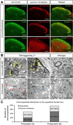
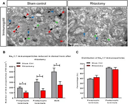
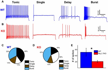
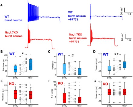
Expression of the voltage-gated sodium channel NaV1.7 in sensory neurons is required for pain sensation. We examined the role of NaV1.7 in the dorsal horn of the spinal cord using an epitope-tagged NaV1.7 knock-in mouse. Immuno-electron microscopy showed the presence of NaV1.7 in dendrites of superficial dorsal horn neurons, despite the absence of mRNA. Rhizotomy of L5 afferent nerves lowered the levels of NaV1.7 in the dorsal horn. Peripheral nervous system-specific NaV1.7 null mutant mice showed central deficits, with lamina II dorsal horn tonic firing neurons more than halved and single spiking neurons more than doubled. NaV1.7 blocker PF05089771 diminished excitability in dorsal horn neurons but had no effect on NaV1.7 null mutant mice. These data demonstrate an unsuspected functional role of primary afferent neuron-generated NaV1.7 in dorsal horn neurons and an expression pattern that would not be predicted by transcriptomic analysis.


Figures

Fig. 1
Fig. 1. The distribution of NaV1.7 in the dorsal horn.
(A) The distribution of TAP-tagged NaV1.7 in the dorsal horn was detected using immunofluorescence with anti-FLAG antibody (left). The cross sections of spinal cord (lumbar 5) were costained with markers of superficial dorsal horn such as substance P (lamina I), PAP (lamina II), and vGLUT1 (lamina III onwards) antibodies (middle). The right panels show the merged left to middle panels. The results show that TAP-tagged NaV1.7 mainly expresses in laminae I and II and in the part of lamina III. Scale bars, 100 μm. (B) Subcellular localization of TAP-tagged NaV1.7 in the dorsal horn of the spinal cord was identified with immuno-EM techniques. Electron micrographs showing immunolabeling for FLAG in the dorsal horn of the spinal cord were detected using the pre-embedding immunoperoxidase (top) and immunogold techniques (bottom). Using the pre-embedding immunoperoxidase method, peroxidase reaction end-product for FLAG in the TAP-tagged NaV1.7 mice was detected filling dendrites (Den) and dendritic spines of spinal cord neurons, as well as at presynaptic sites filling axon terminals (at). In the WT mice (top right), no peroxidase reaction end-product for FLAG was detected in the spinal cord. Using the pre-embedding immunogold method, immunoparticles for FLAG present in the TAP-tagged NaV1.7 were mainly detected at intracellular sites (arrowheads in red), as well as along the plasma membrane (arrowheads in green) in dendritic shafts (Den) of spinal cord neurons. In addition, immunoparticles for FLAG were observed along the extrasynaptic plasma membrane (arrowheads in blue) of axon terminals (at). In the WT mice (bottom right), a very low density of immunoparticles for FLAG, similar to background levels, was observed attached to mitochondria in the spinal cord. Scale bars, 500 nm. HRP, horseradish peroxidase. (C) The number of immunoparticles in the superficial dorsal horn of TAP-tagged NaV1.7 mice was counted. Of 1253 immunoparticles, 768 particles were located at postsynaptic sites (61%) and 485 particles were located at presynaptic sites (39%). Along the 768 postsynaptic particles, 501 particles were located at intracellular sites (65%), and 267 particles were located along the plasma membrane (35%). Along the 485 presynaptic particles, 204 particles were located at intracellular sites (42%), and 281 particles were located along the plasma membrane (58%).
Fig. 2
Fig. 2. TAP-tagged NaV1.7 is down-regulated in the spinal cord after dorsal rhizotomy.
Four weeks after dorsal rhizotomy on the right L5, the spinal cords of TAP-tagged NaV1.7 mice were extracted, and immuno-EM was performed with anti-FLAG antibody and pre-embedding immunogold techniques on the sections adjacent to L5. (A) Representative immuno-EM images from sham control (Sham Ctrl, left) and rhizotomy (right). Immunoparticles for FLAG were detected at intracellular sites (arrowheads in red), plasma membrane (arrowheads in green) in dendritic shafts (Den) of spinal cord neurons, and along the extrasynaptic plasma membrane (arrowheads in blue) of axon terminals (at). Scale bars, 200 nm. (B) The total numbers of immunoparticles from sham control samples (in gray) and rhizotomy (in red) indicate that the expression of TAP-tagged NaV1.7 is down-regulated by about 50% in both presynaptic and postsynaptic terminals after 4 weeks of rhizotomy. (C) Distribution (%) of immunoparticles in both presynaptic and postsynaptic terminals in the rhizotomy model. The numbers of immunoparticles were counted from both presynaptic and postsynaptic terminals. The result shows that there are no significant relative changes of distribution in the rhizotomy model. *P < 0.05, Student’s t test.
Fig. 3
Fig. 3. Electrophysiological properties of lamina II neurons are altered in sensory neuron–specific NaV1.7 KO mice.
Representative traces of the four main firing patterns of lamina II neurons recorded from ex vivo spinal cord slices from (A) WT and (B) NaV1.7 KO mice. Current injections are not shown for clarity. Current injections for recordings shown were as follows: WT tonic = 80 pA, WT single = 200 pA, WT delay = 200 pA, WT burst = 60 pA, KO tonic = 30 pA, KO single = 100 pA, KO delay = 70 pA, and KO burst = 50 pA. Percentage of each neuronal subpopulation in lamina II from (C) WT (n = 83 neurons) and (D) NaV1.7 KO (n = 50 neurons) mice. (E) Maximum number of spikes elicited by current injection for each neuron compared between WT and NaV1.7 KO mice. The difference is statistically significant (*P = 0.02875, two-sample t test with Welch correction) with WT at 51.7 ± 13.2 spikes (n = 83 neurons) and NaV1.7 KO at 21.2 ± 3.8 spikes (n = 50 neurons).
Fig. 4
Fig. 4. Effect of specific NaV1.7 blocker PF771 on lamina II neurons from WT and NaV1.7 KO mice.
Two populations of WT neurons were identified: neurons that displayed an increase in rheobase and neurons that showed little or no change in rheobase in the presence of PF771 (see fig. S4). (A) Representative WT burst firing neuron displaying an effect of PF771 on firing. (B) Representative NaV1.7 KO burst firing neuron displaying no effect of PF771 on firing. Current injections for traces were as follows: WT burst = 30 pA and NaV1.7 KO burst = 50 pA. Current injections were identical before and after drug. (B) WT neurons were split on the basis of rheobase change in the presence of PF771. Paired WT neurons displayed an increase in rheobase with PF771 (hatched bar) versus control (open bar; *P = 0.00586, paired Wilcoxon signed rank test) (C) Maximum number of spikes (at the same current injection before and after drug) was significantly reduced with PF771 versus control (#P = 0.04383, paired Wilcoxon signed rank test) in WT neurons showing an increase in rheobase only. (D) Threshold was significantly increased with PF771 versus control (**P = 0.00178, paired Wilcoxon signed rank test) in WT neurons showing an increase in rheobase only. There were no major population differences in terms of response to PF771 identified in NaV1.7 KO mice dorsal horn neurons. There were no significant changes in (E) rheobase, (F) number of spikes, or (G) threshold of NaV1.7 KO dorsal horn neurons in the presence of PF771 versus control.

References

    1. Nahin R. L., Estimates of pain prevalence and severity in adults: United States, 2012. J. Pain 16, 769–780 (2015). - PMC - PubMed
    1. Habib A. M., Matsuyama A., Okorokov A. L., Santana-Varela S., Bras J. T., Aloisi A. M., Emery E. C., Bogdanov Y. D., Follenfant M., Gossage S. J., Gras M., Humphrey J., Kolesnikov A., Le Cann K., Li S., Minett M. S., Pereira V., Ponsolles C., Sikandar S., Torres J. M., Yamaoka K., Zhao J., Komine Y., Yamamori T., Maniatis N., Panov K. I., Houlden H., Ramirez J. D., Bennett D. L. H., Marsili L., Bachiocco V., Wood J. N., Cox J. J., A novel human pain insensitivity disorder caused by a point mutation in ZFHX2. Brain 141, 365–376 (2018). - PMC - PubMed
    1. Cox J. J., Reimann F., Nicholas A. K., Thornton G., Roberts E., Springell K., Karbani G., Jafri H., Mannan J., Raashid Y., al-Gazali L., Hamamy H., Valente E. M., Gorman S., Williams R., McHale D. P., Wood J. N., Gribble F. M., Woods C. G., An SCN9A channelopathy causes congenital inability to experience pain. Nature 444, 894–898 (2006). - PMC - PubMed
    1. Sexton J. E., Cox J. J., Zhao J., Wood J. N., The genetics of pain: Implications for therapeutics. Annu. Rev. Pharmacol. Toxicol. 58, 123–142 (2018). - PubMed
    1. Habib A. M., Wood J. N., Cox J. J., Sodium channels and pain. Handb. Exp. Pharmacol. 227, 39–56 (2015). - PubMed

Publication types

MeSH terms

Substances