. 2020 Feb 19;6(8):eaay6812.
doi: 10.1126/sciadv.aay6812. eCollection 2020 Feb.

Enhanced CRISPR-Cas9 correction of Duchenne muscular dystrophy in mice by a self-complementary AAV delivery system

Affiliations

Enhanced CRISPR-Cas9 correction of Duchenne muscular dystrophy in mice by a self-complementary AAV delivery system

Yu Zhang et al. Sci Adv. .

Abstract

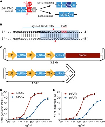
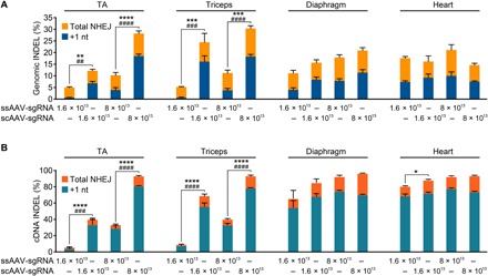
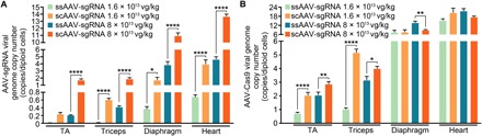
Duchenne muscular dystrophy (DMD) is a lethal neuromuscular disease caused by mutations in the dystrophin gene (DMD). Previously, we applied CRISPR-Cas9-mediated "single-cut" genome editing to correct diverse genetic mutations in animal models of DMD. However, high doses of adeno-associated virus (AAV) are required for efficient in vivo genome editing, posing challenges for clinical application. In this study, we packaged Cas9 nuclease in single-stranded AAV (ssAAV) and CRISPR single guide RNAs in self-complementary AAV (scAAV) and delivered this dual AAV system into a mouse model of DMD. The dose of scAAV required for efficient genome editing were at least 20-fold lower than with ssAAV. Mice receiving systemic treatment showed restoration of dystrophin expression and improved muscle contractility. These findings show that the efficiency of CRISPR-Cas9-mediated genome editing can be substantially improved by using the scAAV system. This represents an important advancement toward therapeutic translation of genome editing for DMD.


Figures

Fig. 1
Fig. 1. Strategies for CRISPR-Cas9–mediated genome editing in Dmd ∆Ex44 mice.
(A) An out-of-frame deletion of Dmd exon 44 results in splicing of exons 43 to 45, generating a premature stop codon in exon 45. A CRISPR-Cas9–mediated “single-cut” strategy was designed to restore the open reading frame (ORF) of the Dmd gene. If the genomic insertions and deletions (INDELs) result in one nucleotide insertion (3n + 1) or two nucleotide deletion (3n − 2), then exon 45 will be reframed with adjacent exons 43 and 46. If the INDEL is large enough to delete the 5′-AG-3′ splice acceptor sequence, then exon 45 will be skipped, resulting in splicing of exon 43 to exon 46. (B) Illustration of sgRNA targeting Dmd exon 45. This sgRNA recognizes a 5′-TGG-3′ PAM in exon 45 and generates a cut 7 base pairs downstream of the 5′-AG-3′ splice acceptor site. (C) Illustration of AAV vectors used to deliver the sgRNA expression cassette. Three copies of the same sgRNA are driven by three RNA polymerase III promoters, U6, H1, and 7SK. The top vector produces ssAAV. A 2.3-kb stuffer sequence was cloned into the ssAAV vector for optimal packaging. The bottom vector produces double-stranded scAAV. (D) Analysis of total INDEL event in 5-day differentiated myotubes transduced with scAAV- or ssAAV-packaged sgRNA at multiple doses. Data are presented as mean ± SEM (n = 3). (E) Analysis of the +1-nt insertion event in 5-day differentiated myotubes transduced with scAAV- or ssAAV-packaged sgRNA at multiple doses. Data are presented as mean ± SEM (n = 3).
Fig. 2
Fig. 2. Systemic AAV delivery of CRISPR-Cas9 genome editing components to ∆Ex44 mice rescues dystrophin expression.
Immunohistochemistry shows restoration of dystrophin in the tibialis anterior (TA), triceps, diaphragm, and heart of ∆Ex44 mice 4 weeks after systemic delivery of ssAAV-packaged SpCas9 and scAAV-packaged sgRNA. The SpCas9 vector was kept at a constant dose of 8 × 10¹³ vg/kg. The dose of the sgRNA vector is shown in the figure. Dystrophin is shown in green. n = 5 for each muscle group. Scale bars, 100 μm.
Fig. 3
Fig. 3. Western blot analysis of skeletal muscles and heart of ∆Ex44 mice receiving systemic AAV delivery of CRISPR-Cas9 genome editing components.
(A) Western blot analysis shows restoration of dystrophin expression in the TA, triceps, diaphragm, and heart of ∆Ex44 mice 4 weeks after systemic delivery of ssAAV-packaged SpCas9 and scAAV-packaged sgRNA. The SpCas9 vector was kept at a constant dose of 8 × 10¹³ vg/kg. The dose of the sgRNA vector is shown in the figure. Vinculin was used as the loading control (n = 3). (B) Quantification of dystrophin expression in the TA, triceps, diaphragm, and heart. Relative dystrophin intensity was calibrated with vinculin internal control before normalizing to the WT control. Data are presented as mean ± SEM. One-way ANOVA was performed with post hoc Tukey’s multiple comparisons test. **P < 0.005, ***P < 0.001, ****P < 0.0001 (n = 3). (C) Quantification of Cas9 expression in the TA, triceps, diaphragm, and heart. Relative Cas9 intensity was calibrated with vinculin internal control before normalizing to the group treated with the lowest dose of scAAV-packaged sgRNA (4 × 10¹² vg/kg). Data are presented as mean ± SEM. One-way ANOVA was performed with post hoc Tukey’s multiple comparisons test. *P < 0.05, **P < 0.005 (n = 3).
Fig. 4
Fig. 4. Rescue of skeletal muscle function after systemic AAV delivery of CRISPR-Cas9 genome editing components.
(A and B) Specific force (mN/mm2) of the extensor digitorum longus (EDL) (A) and soleus (B) muscles in WT, ∆Ex44 mice untreated, and ∆Ex44 mice treated with ssAAV-packaged SpCas9 and scAAV- or ssAAV-packaged sgRNA. The SpCas9 vector was kept at a constant dose of 8 × 10¹³ vg/kg. The dose of the sgRNA vector is shown in the figure. Data are presented as mean ± SEM. One-way ANOVA was performed with post hoc Tukey’s multiple comparisons test. *P < 0.05, **P < 0.005, ***P < 0.001, ****P < 0.0001 (n = 6). (C and D) Maximal tetanic force of the EDL (C) and soleus (D) muscles in WT, ∆Ex44 untreated mice, and ∆Ex44 mice treated with ssAAV-packaged SpCas9 and scAAV- or ssAAV-packaged sgRNA. The SpCas9 vector was kept at a constant dose of 8 × 10¹³ vg/kg. The dose of the sgRNA vector is shown in the figure (n = 6).
Fig. 5
Fig. 5. The scAAV vector induces significant INDELs at the genomic and cDNA level.
(A and B) Genomic INDEL analysis by deep sequencing (A) and dystrophin cDNA INDEL analysis by TIDE analysis (B) of the TA, triceps, diaphragm, and heart of ∆Ex44 mice 4 weeks after systemic delivery of ssAAV-packaged SpCas9 and scAAV or ssAAV-packaged sgRNA. The SpCas9 vector was kept at a constant dose of 8 × 10¹³ vg/kg. The dose of the sgRNA vector is shown in the figure. Data are presented as mean ± SEM. Two-way ANOVA was performed with post hoc Tukey’s multiple comparisons test. *P < 0.05, **P < 0.005, ***P < 0.001, ****P < 0.0001 for the total NHEJ event (n = 3). ##P < 0.005, ###P < 0.001, ####P < 0.0001 for the +1-nt insertion event (n = 3).
Fig. 6
Fig. 6. ∆Ex44 mice sustain higher copies of viral genome after systemic delivery of scAAV-packaged sgRNA.
(A and B) sgRNA viral genome copy number (A) and Cas9 viral genome copy number (B) quantification from skeletal muscles and heart of ∆Ex44 mice 4 weeks after systemic delivery of ssAAV-packaged SpCas9 and scAAV- or ssAAV-packaged sgRNA. The SpCas9 vector was kept at a constant dose of 8 × 10¹³ vg/kg. The dose of the sgRNA vector is shown in the figure. Data are presented as mean ± SEM. One-way ANOVA was performed with post hoc Tukey’s multiple comparisons test. *P < 0.05, **P < 0.005, ****P < 0.0001 (n = 3).

Similar articles

Cited by

References

    1. Hoffman E. P., Brown R. H. Jr., Kunkel L. M., Dystrophin: The protein product of the Duchenne muscular dystrophy locus. Cell 51, 919–928 (1987). - PubMed
    1. Koenig M., Hoffman E. P., Bertelson C. J., Monaco A. P., Feener C., Kunkel L. M., Complete cloning of the Duchenne muscular dystrophy (DMD) cDNA and preliminary genomic organization of the DMD gene in normal and affected individuals. Cell 50, 509–517 (1987). - PubMed
    1. Gao Q., McNally E. M., The dystrophin complex: Structure, function, and implications for therapy. Compr. Physiol. 5, 1223–1239 (2015). - PMC - PubMed
    1. Guiraud S., Aartsma-Rus A., Vieira N. M., Davies K. E., van Ommen G.-J. B., Kunkel L. M., The pathogenesis and therapy of muscular dystrophies. Annu. Rev. Genomics Hum. Genet. 16, 281–308 (2015). - PubMed
    1. Campbell K. P., Kahl S. D., Association of dystrophin and an integral membrane glycoprotein. Nature 338, 259–262 (1989). - PubMed

Publication types

MeSH terms