. 2020 Jan 13;5(1):157-162.
doi: 10.1002/lio2.347. eCollection 2020 Feb.

Otopathology in CHARGE syndrome

Affiliations

Otopathology in CHARGE syndrome

Jenny X Chen et al. Laryngoscope Investig Otolaryngol. .

Abstract

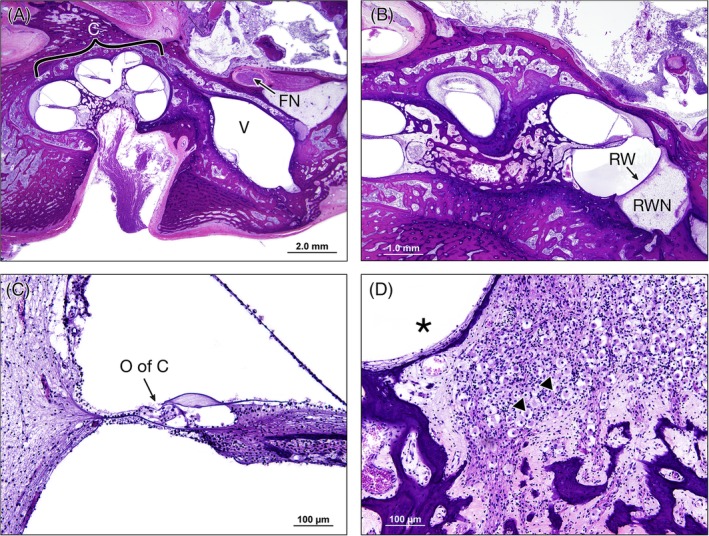
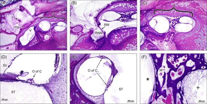
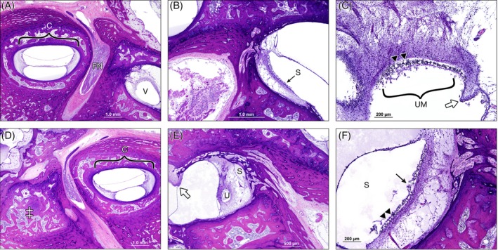
Postmortem temporal bone computed tomography (CT) and histopathologic findings in an infant with CHARGE syndrome revealed bilateral cochleovestibular hypoplasia, including cochlear pathology relevant to cochlear implant candidacy. Both ears had absence of the superior semicircular canals (SCCs), severely hypoplastic posterior SCCs, and hypoplastic (right ear) or absent (left ear) lateral SCCs seen on CT and histopathology. Histopathology further revealed the absence of all SCC ampullae except the right lateral SCC ampulla and atrophic vestibular neuroepithelium in the saccule and utricle bilaterally. The right cochlea consisted of a basal turn with patent round window, and malformed middle turn (type IV cochlear hypoplasia), with a small internal auditory canal (IAC) but near normal cochlear nerve aperture (fossette). Quantification of spiral ganglion neurons (SGNs) on histologic sections revealed a reduced SGN population (35% of normal for age), but this ear would still have likely achieved benefit from a cochlear implant based on this population. The left cochlea consisted of only a basal turn with patent round window (type III cochlear hypoplasia) with a small IAC and very small cochlear nerve aperture. Notably, histology revealed that there were no SGNs in the cochlea, and therefore, this ear would not have been a good candidate for cochlear implantation.

Level of evidence: IV.

Keywords: CHARGE syndrome; cochlear implantation; congenital anomalies; otopathology; temporal bone pathology.


Conflict of interest statement

None.

Figures

Figure 1
Figure 1
Axial CT of the right and left temporal bones. A, Right hypoplastic cochlea with basal turn (thick arrow) and underdeveloped middle turn (thin arrow). The cochlear aperture is near normal caliber (dotted arrows). Malleus (M) and incus (I) present in the middle ear. B, Right vestibule (V) present. Note, the finger‐like projection off the vestibule is the take off of the hypoplastic posterior semicircular canal. C, Left hypoplastic cochlea with basal turn (thick arrows) and a very small cochlear nerve aperture (dotted arrows). D, Left vestibule (V) present with hypoplastic posterior semicircular canal
Figure 2
Figure 2
Cochlear pathology in the right ear. A, Low magnification near mid‐modiolar section showing foreshortened cochlea (C) with two turns. There is no oval window or stapes and thus the facial nerve (FN) crosses directly over the vestibule (V), 1.25×. B, Normally formed round window (RW) and patent round window niche (RWN), 2×. C, Lower basal turn of the cochlea showing normal complement of outer hair cells in the organ of Corti (O of C) and some dendritic fibers of spiral ganglion cells, 10×. D, Mid‐modiolar section next to the scala tympani of the middle turn (*) showing significantly reduced numbers of spiral ganglion cells (arrow heads), 10×
Figure 3
Figure 3
Cochlear pathology in the left ear. A, Low magnification view of the middle ear with the lenticular process of the incus (I) as well as the facial nerve (FN) coursing over the vestibule (V), 1.25×. Basal turn of the cochlea (c). B, The round window (RW) and round window niche (RWN) are normally formed. C, The cochlea (c) is hypoplastic with missing turns, a very small fundus and cochlear nerve aperture, 2×. D, The lower basal turn of the cochlea with proteinaceous debris in the scala vestibule (SV) and scala tympani (ST). The osseous spiral lamina appears empty. The organ of corti appears fully formed. E, In the upper basal turn, there is greater deformity of the cochlea as shown by the development of two organ of Corti (O of C) and tectorial membranes, 10×. F, Modiolus (M) between the scala tympani of the upper (*) and lower (+) basal turns with no spiral ganglion cells, 10×
Figure 4
Figure 4
Vestibular findings. Right ear: A, Abnormal position of the geniculate ganglion cells located within the labyrinthine facial nerve (FN), 2×. Vestibule (V) is present but there is no superior SCC. Basal turn of hypoplastic cochlea (c). B, Small saccule (S) with vestibular neuroepithelium (arrow). C, Utricular macula (UM) with vacuolated spaces consistent with the presence of type 1 vestibular hair cells (arrow heads). The vestigial ampulla of the lateral SCC (open arrow) with limited true neuroepithelium, 10×. Left ear: D, Significant vestibular hypoplasia notable for lack of superior SCC typically in the area marked (‡), 2×. E, The beginning of the lateral SCCa ampulla (open arrow), with underdeveloped utricle (U) and saccule (S), 4×. F, Saccule (S) with neuroepithelium present (arrow) and vacuolated spaces consistent with the presence of type 1 vestibular hair cells (arrow heads), 10×

Similar articles

Cited by

References

    1. Vesseur AC, Verbist BM, Westerlaan HE, et al. CT findings of the temporal bone in CHARGE syndrome: aspects of importance in cochlear implant surgery. Eur Arch Otorhinolaryngol. 2016;273(12):4225‐4240. 10.1007/s00405-016-4141-z. - DOI - PMC - PubMed
    1. Merchant S, Nadol J. Methods of removal, preparation and study Schuknecht's Pathology of the Ear. Shelton, CT: People's Medical Pub. House‐USA Inc; 2010:3Y51.
    1. Sennaroğlu L, Demir Bajin M. Classification and current management of inner ear malformations. Balkan Med J. 2017;34(5):397‐411. 10.4274/balkanmedj.2017.0367. - DOI - PMC - PubMed
    1. Haginomori S‐I, Miura M, Sando I, Casselbrant ML. Temporal bone histopathology in CHARGE association. Ann Otol Rhinol Laryngol. 2002;111(5):397‐401. 10.1177/000348940211100503. - DOI - PubMed
    1. Young NM, Tournis E, Sandy J, Hoff SR, Ryan M. Outcomes and time to emergence of auditory skills after cochlear implantation of children with CHARGE syndrome. Otol Neurotol. 2017;38(8):1085‐1091. 10.1097/MAO.0000000000001488. - DOI - PubMed

LinkOut - more resources