

Precision wearable accelerometer contact microphones for longitudinal monitoring of mechano-acoustic cardiopulmonary signals
- PMID: 32128449
- PMCID: PMC7015926
- DOI: 10.1038/s41746-020-0225-7
Precision wearable accelerometer contact microphones for longitudinal monitoring of mechano-acoustic cardiopulmonary signals
Abstract
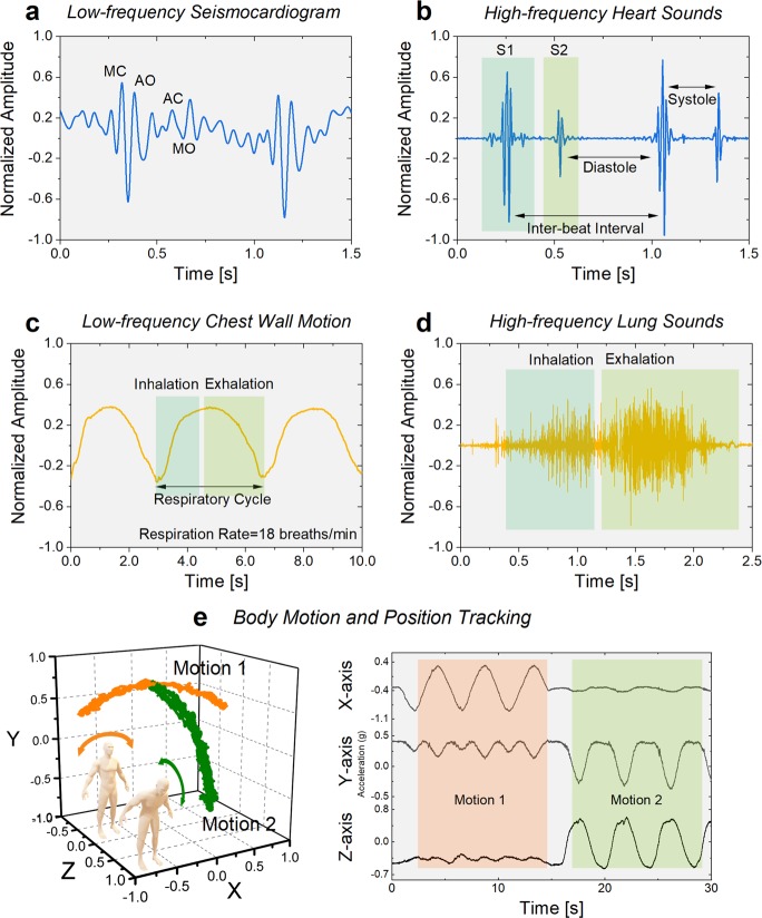
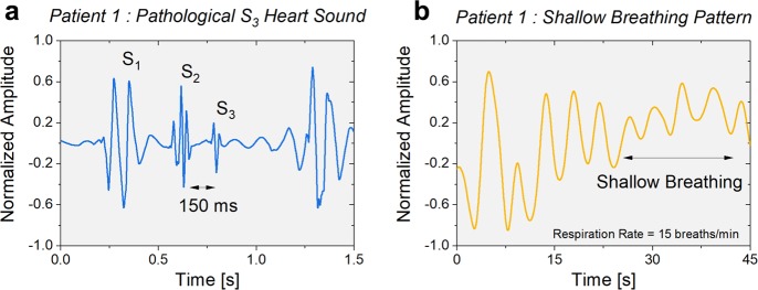
Mechano-acoustic signals emanating from the heart and lungs contain valuable information about the cardiopulmonary system. Unobtrusive wearable sensors capable of monitoring these signals longitudinally can detect early pathological signatures and titrate care accordingly. Here, we present a wearable, hermetically-sealed high-precision vibration sensor that combines the characteristics of an accelerometer and a contact microphone to acquire wideband mechano-acoustic physiological signals, and enable simultaneous monitoring of multiple health factors associated with the cardiopulmonary system including heart and respiratory rate, heart sounds, lung sounds, and body motion and position of an individual. The encapsulated accelerometer contact microphone (ACM) utilizes nano-gap transducers to achieve extraordinary sensitivity in a wide bandwidth (DC-12 kHz) with high dynamic range. The sensors were used to obtain health factors of six control subjects with varying body mass index, and their feasibility in detection of weak mechano-acoustic signals such as pathological heart sounds and shallow breathing patterns is evaluated on patients with preexisting conditions.
Keywords: Biomedical engineering; Electrical and electronic engineering; Health care.
© The Author(s) 2020.
Conflict of interest statement
Competing interestsP.G., Y.J. and F.A. are the inventors of the technology being studied and the purpose of this project is to explore its commercialization. The terms of arrangement have been reviewed and approved by Georgia Tech in accordance with its conflict of interest policies. O.T.I. is a scientific advisor for Physiowave Inc., a manufacturer of ballistocardiogram sensing hardware. M.J.M. and D.G. declare that there are no competing interests.
Figures


Similar articles
-
Detection of pathological mechano-acoustic signatures using precision accelerometer contact microphones in patients with pulmonary disorders.Sci Rep. 2021 Jun 28;11(1):13427. doi: 10.1038/s41598-021-92666-2. Sci Rep. 2021. PMID: 34183695 Free PMC article.
-
An Accelerometer-Based Wearable Patch for Robust Respiratory Rate and Wheeze Detection Using Deep Learning.Biosensors (Basel). 2024 Feb 22;14(3):118. doi: 10.3390/bios14030118. Biosensors (Basel). 2024. PMID: 38534225 Free PMC article.
-
Algorithm for heart rate extraction in a novel wearable acoustic sensor.Healthc Technol Lett. 2015 Feb 24;2(1):28-33. doi: 10.1049/htl.2014.0095. eCollection 2015 Feb. Healthc Technol Lett. 2015. PMID: 26609401 Free PMC article.
-
Physiological acoustic sensing based on accelerometers: a survey for mobile healthcare.Ann Biomed Eng. 2014 Nov;42(11):2264-77. doi: 10.1007/s10439-014-1111-8. Epub 2014 Sep 19. Ann Biomed Eng. 2014. PMID: 25234130 Review.
-
Advances in Microsensors and Wearable Bioelectronics for Digital Stethoscopes in Health Monitoring and Disease Diagnosis.Adv Healthc Mater. 2021 Nov;10(22):e2101400. doi: 10.1002/adhm.202101400. Epub 2021 Sep 5. Adv Healthc Mater. 2021. PMID: 34486237 Review.
Cited by
-
Exploring Microphone Technologies for Digital Auscultation Devices.Micromachines (Basel). 2023 Nov 12;14(11):2092. doi: 10.3390/mi14112092. Micromachines (Basel). 2023. PMID: 38004949 Free PMC article.
-
Electrostatic Acoustic Sensor with an Impedance-Matched Diaphragm Characterized for Body Sound Monitoring.ACS Appl Bio Mater. 2023 Aug 21;6(8):3241-3256. doi: 10.1021/acsabm.3c00359. Epub 2023 Jul 20. ACS Appl Bio Mater. 2023. PMID: 37470762 Free PMC article.
-
Sensing Devices for Detecting and Processing Acoustic Signals in Healthcare.Biosensors (Basel). 2022 Oct 7;12(10):835. doi: 10.3390/bios12100835. Biosensors (Basel). 2022. PMID: 36290973 Free PMC article. Review.
-
Sensory Stimulation in the NICU Environment: Devices, Systems, and Procedures to Protect and Stimulate Premature Babies.Children (Basel). 2021 Apr 25;8(5):334. doi: 10.3390/children8050334. Children (Basel). 2021. PMID: 33923031 Free PMC article. Review.
-
Integration of novel monitoring devices with machine learning technology for scalable cardiovascular management.Nat Rev Cardiol. 2021 Feb;18(2):75-91. doi: 10.1038/s41569-020-00445-9. Epub 2020 Oct 9. Nat Rev Cardiol. 2021. PMID: 33037325 Free PMC article. Review.
References
Grants and funding
LinkOut - more resources
Full Text Sources
Other Literature Sources
Miscellaneous
