. 2020 May;111(5):1582-1595.
doi: 10.1111/cas.14372. Epub 2020 Apr 30.

Long noncoding RNA metastasis-associated lung adenocarcinoma transcript 1 cooperates with enhancer of zeste homolog 2 to promote hepatocellular carcinoma development by modulating the microRNA-22/Snail family transcriptional repressor 1 axis

Affiliations

Long noncoding RNA metastasis-associated lung adenocarcinoma transcript 1 cooperates with enhancer of zeste homolog 2 to promote hepatocellular carcinoma development by modulating the microRNA-22/Snail family transcriptional repressor 1 axis

Shaofei Chen et al. Cancer Sci. 2020 May.

Abstract

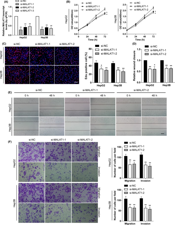
Metastasis-associated lung adenocarcinoma transcript 1 (MALAT1) is an oncogenic long noncoding RNA that has been found to promote carcinogenesis and metastasis in many tumors. However, the underlying role of MALAT1 in the progression and metastasis of hepatocellular carcinoma (HCC) remains unclear. In this study, aberrantly elevated levels of MALAT1 were detected in both HCC specimens and cell lines. We found that knockdown of MALAT1 caused retardation in proliferation, migration, and invasion both in vivo and in vitro. Mechanistic investigations showed that Snail family transcriptional repressor 1 (SNAI1) is a direct target of microRNA (miR)-22 and that MALAT1 modulates SNAI1 expression by acting as a competing endogenous RNA for miR-22. Inhibition of miR-22 restored SNAI1 expression suppressed by MALAT1 knockdown. Furthermore, MALAT1 facilitated the enrichment of enhancer of zeste homolog 2 (EZH2) at the promoter region of miR-22 and E-cadherin, which was repressed by MALAT1 knockdown. Cooperating with EZH2, MALAT1 positively regulated SNAI1 by repressing miR-22 and inhibiting E-cadherin expression, playing a vital role in epithelial to mesenchymal transition. In conclusion, our results reveal a mechanism by which MALAT1 promotes HCC progression and provides a potential target for HCC therapy.

Keywords: EZH2; MALAT1; SNAI1; hepatocellular carcinoma; miR-22.


Conflict of interest statement

The authors have no conflict of interest for this article.

Figures

Figure 1
Figure 1
Metastasis‐associated lung adenocarcinoma transcript 1 (MALAT1) is upregulated in hepatocellular carcinoma (HCC) tissues and cell lines and negatively correlated with microRNA (miR)‐22. A, MALAT1 expression was determined by quantitative PCR and normalized to GAPDH in 30 paired HCC tissues and adjacent noncancerous tissues. B, MALAT1 expression levels of HCC and paired normal tissues were analyzed and expressed as log2 fold change (HCC/normal), and the log2 fold changes were presented as follows: >1, overexpression (17 cases); <−1, underexpression (3 cases); and the remainder were defined as unchanged (10 cases). C, HCC cell lines expressed higher levels of MALAT1 and lower levels of miR‐22 than normal liver tissues. D, Higher MALAT1 and lower miR‐22 levels were detected in high‐stage patients than low‐stage patients. E, Box plot representation of MALAT1 levels in normal liver (n = 10) and HCC (n = 35) samples. Analysis is based on RNA sequencing data from the Oncomine database (https://www.oncomine.org). F, G, Associations of MALAT1 (F) and miR‐22 (G) expression with overall survival were analyzed by Kaplan‐Meier plots. Survival data were based on The Cancer Genome Atlas (TCGA) datasets and analyzed by SurvExpress (http://bioinformatica.mty.itesm.mx/SurvExpress) and UCSC Xena (http://xena.ucsc.edu/), respectively. H, I, Inverse correlations were found between MALAT1 and miR‐22 in both clinical specimens (H) and TCGA datasets (I). (*P < .05, **P < .01)
Figure 2
Figure 2
Knockdown of metastasis‐associated lung adenocarcinoma transcript 1 (MALAT1) inhibits cell proliferation, migration, and invasion of hepatocellular carcinoma (HCC) cells in vitro. A, MALAT1 levels were detected after transfection with siRNAs specific for MALAT1 in HepG2 and Hep3B cells. The most effective 2 were selected for further study. B, CCK‐8 assays showed that knockdown of MALAT1 decreased the proliferation abilities of HCC cells. C, Edu assays showed that suppression of MALAT1 attenuated the proliferation rate of HCC cells (scale bar, 50 µm). D, E, Wound healing assays showed that MALAT1 knockdown resulted in a delayed closure of scratch wounds (scale bar, 100 µm). F, Transwell migration and invasion assays showed that knockdown of MALAT1 attenuated the migration and invasion abilities of HCC cells (scale bar, 50 µm). *P < .05, **P < .01, compared with control (si‐NC)
Figure 3
Figure 3
Metastasis‐associated lung adenocarcinoma transcript 1 (MALAT1) regulates microRNA (miR)‐22 expression by directly binding to miR‐22 in hepatocellular carcinoma (HCC) cells. A, Schematic representation of the putative binding sites of miR‐22 on the MALAT1 transcript, as predicted by LncBase Predicted version 2. B, Expression levels of MALAT1 in HCC cells transfected with miR‐22 mimic or inhibitor. C, miR‐22 levels were detected after knockdown and overexpression MALAT1, respectively. D, RNA immunoprecipitation assays were carried out using anti‐Ago2 Ab. Relative expression levels of MALAT1 and miR‐22 in HCC cells were detected by quantitative PCR (qPCR). Expressions of MALAT1 and miR‐22 were normalized by GAPDH and U6, respectively. E, HCC cell lysates were incubated with biotinylated MALAT1 probe containing the putative binding sites of miR‐22 or the biotinylated antisense strand. Lysates incubated with beads only were used as negative control (NC), input was used for normalization. MiR‐22 levels were analyzed by qPCR. F, Luciferase activities of HCC cells were detected after cotransfection with indicated vectors and miR‐22 mimic or control. *P < .05, **P < .01, compared with respective control
Figure 4
Figure 4
Metastasis‐associated lung adenocarcinoma transcript 1 (MALAT1) interacts with enhancer of zeste homolog 2 (EZH2) in hepatocellular carcinoma. A, RNA pull‐down assays were carried out with biotinylated MALAT1 probe followed by western blot assay using anti‐EZH2 Ab. B, Schematic illustration of MALAT1 regions used for truncated RNA pull‐down assays. C, RNA pull‐down assays using biotinylated truncated MALAT1 confirmed that MALAT1 bound EZH2 mainly at its 3′‐end. D, Enrichment of the MALAT1 fractions in cross‐link and sonication RNA immunoprecipitation (RIP) using anti‐EZH2 Ab further validated EZH2 interacted with the 3′‐end of MALAT1. E, Anti‐EZH2 RIP was carried out after transiently overexpressing microRNA (miR)‐22 followed by detecting MALAT1 with quantitative PCR. miR‐22 did not compete with EZH2 for binding MALAT1. F, G, ChIP assays were undertaken with anti‐EZH2 and anti‐H3K27me3 Abs after knockdown of MALAT1. Knockdown of MALAT1 attenuated the enrichment of EZH2 at miR‐22 (F) and E‐cadherin (G) promoter. *P < .05, **P < .01, compared with respective control
Figure 5
Figure 5
Snail family transcriptional repressor 1 (SNAI1) is a direct target of microRNA (miR)‐22 in hepatocellular carcinoma (HCC). A, Schematic representation and sequence of the intact miR‐22 binding site (WT) and its mutation form (MUT) within the luciferase reporter vector. B, HCC cells were cotransfected with indicated vector and miR‐22 mimic or control (NC), luciferase activities were determined 48 h after transfection. C, HCC cells were transfected with miR‐22 mimic (miR‐22 mmc) or inhibitor (anti‐miR‐22), followed by western blot assays to identify expression of SNAI1. D, Relative luciferase activities of HCC cells transfected with SNAI1 3′‐UTR WT vectors were determined after knockdown or upregulation of metastasis‐associated lung adenocarcinoma transcript 1 (MALAT1). E, F, SNAI1 expression levels in HCC cells were determined by western blot assays after knockdown or upregulation of MALAT1. G, A negative correlation was found between miR‐22 and SNAI1 mRNA levels in clinical specimens. H, MALAT1 was positively correlated with SNAI1 levels, confirmed by quantitative PCR in HCC tissues. *P < .05, **P < .01, compared with respective control
Figure 6
Figure 6
Decreased abilities of proliferation, migration, and invasion caused by metastasis‐associated lung adenocarcinoma transcript 1 (MALAT1) knockdown were partially reversed by microRNA (miR)‐22 inhibition in vitro. Hepatocellular carcinoma (HCC) cells stably transfected with empty vector (mock) or sh‐MALAT1 were cotransfected with negative control inhibitor (anti‐ctrl, 100 nmol/L) or anti‐miR‐22 inhibitor (100 nmol/L). A, Western blot assays of epithelial‐mesenchymal transition markers (ie, E‐cadherin, vimentin, or Snail family transcriptional repressor 1 [SNAI1]) indicated that altered expressions caused by MALAT1 knockdown were reversed by miR‐22 inhibition. B, Representative images (upper) and quantification (lower) of Edu proliferation assays showed that inhibition of miR‐22 restored the proliferation abilities of HCC cells decreased by MALAT1 knockdown (scale bar, 50 µm). C, Representation (left panel) and quantification (right panel) of colony formation assays showed the clonogenicity abilities of HCC cells transfected with indicated vectors (scale bar, 1 cm). D, Knockdown of MALAT1 attenuated the migration and invasion of HCC cells, which was reversed by transfection of anti‐miR‐22 (scale bar, 50 µm). *P < .05, **P < .01, compared with respective control
Figure 7
Figure 7
Knockdown of metastasis‐associated lung adenocarcinoma transcript 1 (MALAT1) inhibited tumor growth and metastasis in vivo, and it could be restored by inhibition of microRNA (miR)‐22. HepG2 cells stably transfected with empty vector (mock) or sh‐MALAT1 were cotransfected with negative control inhibitor (anti‐ctrl, 100 nmol/L) or anti‐miR‐22 inhibitor (100 nmol/L). A, Representative fluorescence images of mice (n = 5 each group) injected with HepG2 cells stably transfected with indicated plasmids bearing the cherry fluorescent protein. Bar graph showing the quantification of normalized total photon counts of s.c. xenografts in mice of each group. B, In vivo growth curves of xenograft tumors after hypodermic injection of indicated HepG2 cells. C, Representation (upper panel) and quantification (lower panel) of xenograft tumors formed by hypodermic injection of HepG2 cells transfected with indicated vectors. D, Immunohistochemical staining for Snail family transcriptional repressor 1 (SNAI1) and E‐cadherin expression within tumors formed by hypodermic injections of HepG2 cells stably transfected with indicated vectors (scale bar, 100 µm). E, Representation (left panels, arrowhead; scale bar, 500 µm) and quantification (right panels; scale bar, 100 µm) of lung metastasis of nude mice injected with HepG2 cells (2 × 106) stably transfected with mock or sh‐MALAT1, and those cotransfected with anti‐miR‐22 or control through the tail vein (n = 5, each group)

References

    1. El‐Serag HB, Rudolph KL. Hepatocellular carcinoma: epidemiology and molecular carcinogenesis. Gastroenterology. 2007;132:2557‐2576. - PubMed
    1. Li H, Wu K, Tao K, et al. Tim‐3/galectin‐9 signaling pathway mediates T‐cell dysfunction and predicts poor prognosis in patients with hepatitis B virus‐associated hepatocellular carcinoma. Hepatology. 2012;56:1342‐1351. - PubMed
    1. Chen W, Zheng R, Baade PD, et al. Cancer statistics in China, 2015. CA Cancer J Clin. 2016;66:115‐132. - PubMed
    1. Siegel RL, Miller KD, Jemal A. Cancer statistics, 2016. CA Cancer J Clin. 2016;66(1):7–30. - PubMed
    1. Djebali S, Davis CA, Merkel A, et al. Landscape of transcription in human cells. Nature. 2012;489:101‐108. - PMC - PubMed

MeSH terms