. 2020 Mar 4;16(3):e1008604.
doi: 10.1371/journal.pgen.1008604. eCollection 2020 Mar.

Murine cytomegalovirus infection exacerbates complex IV deficiency in a model of mitochondrial disease

Affiliations

Murine cytomegalovirus infection exacerbates complex IV deficiency in a model of mitochondrial disease

Nicola Ferreira et al. PLoS Genet. .

Abstract

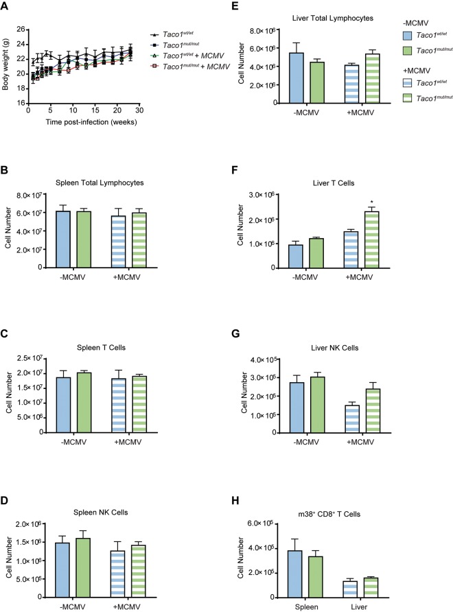
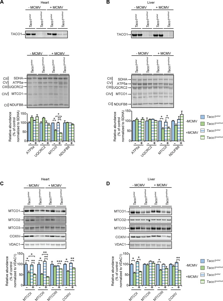
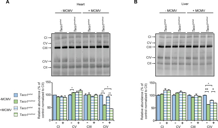
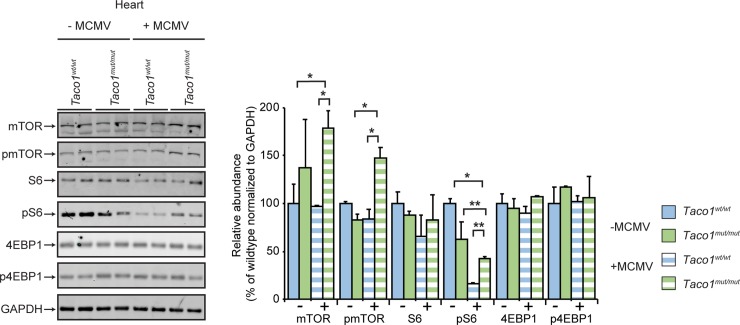
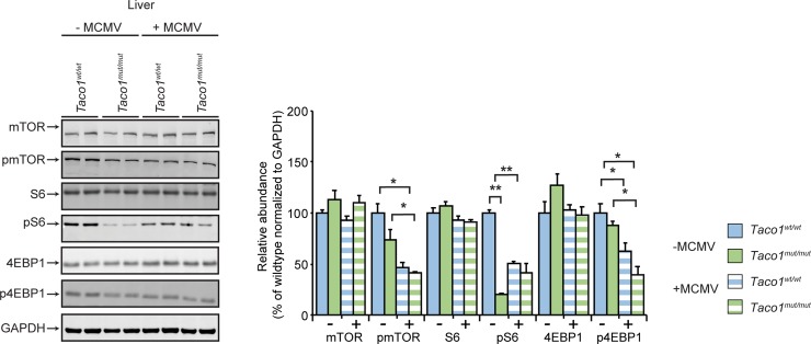
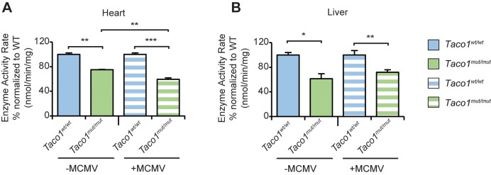
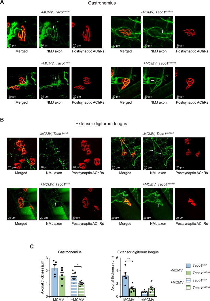
The influence of environmental insults on the onset and progression of mitochondrial diseases is unknown. To evaluate the effects of infection on mitochondrial disease we used a mouse model of Leigh Syndrome, where a missense mutation in the Taco1 gene results in the loss of the translation activator of cytochrome c oxidase subunit I (TACO1) protein. The mutation leads to an isolated complex IV deficiency that mimics the disease pathology observed in human patients with TACO1 mutations. We infected Taco1 mutant and wild-type mice with a murine cytomegalovirus and show that a common viral infection exacerbates the complex IV deficiency in a tissue-specific manner. We identified changes in neuromuscular morphology and tissue-specific regulation of the mammalian target of rapamycin pathway in response to viral infection. Taken together, we report for the first time that a common stress condition, such as viral infection, can exacerbate mitochondrial dysfunction in a genetic model of mitochondrial disease.


Conflict of interest statement

The authors have declared that no competing interests exist.

Figures

Fig 1
Fig 1. Taco1 mut/mut mice generate a normal immune response.
(A) Body weights from uninfected and MCMV infected Taco1wt/wt and Taco1 mut/mut mice. Spleens and livers from uninfected and MCMV infected 30 week old Taco1wt/wt and Taco1 mut/mut mice were prepared for flow cytometry to determine (B) the number of total spleen lymphocytes, (C) the number of spleen T cells, (D) the number of spleen NK cells, (E) the number of total liver lymphocytes, (F) the number of liver T cells, (G) the number of liver NK cells, and (H) the number of m38+ CD8+ T cells in the spleen and liver. The data are representative of results obtained from at least 5 mice from each genotype and each infection group. Error bars indicate SEM; *p<0.05, compared with control treatments by a two-tailed paired Student’s t-test.
Fig 2
Fig 2. MCMV infection reduces the abundance of complex IV subunits in the heart.
Homogenates from uninfected and MCMV infected 30 week old Taco1wt/wt mice and Taco1 mut/mut adult mice were resolved on 4–12% Bis-Tris gels and immunoblotted using antibodies to investigate the steady state levels of TACO1 and OXPHOS protein subunits in the heart (25 μg) (A) and in the liver (50 μg) (B), as well as mitochondrial and nuclear-encoded Complex IV protein subunits in the heart (C) and in the liver (D). CII (Succinate dehydrogenase complex subunit A (SDHA)) or VDAC1 were used as loading controls. Relative abundance of proteins was measured using Li-Cor Odyssey Classic software normalized to the loading control. All data are representative of results obtained from 4 mice of each strain and two independent biological experiments. Error bars indicate SEM; *p<0.05, **p<0.01, compared with control treatments by a two-tailed paired Student’s t-test.
Fig 3
Fig 3. MCMV infection exacerbates mitochondrial dysfunction in the heart.
(A) Heart homogenates (80 μg) and (B) liver homogenates (160 μg) from uninfected and MCMV infected 30 week old Taco1wt/wt mice and Taco1 mut/mut adult mice were resolved on a 4–16% BN-PAGE gels. Immunoblotting with the Blue Native OXPHOS antibody cocktail was used to visualize respiratory complexes. CII was used as a loading control. Relative abundance of proteins was measured using Li-Cor Odyssey Classic software normalized to the loading control. All data are representative of results obtained from 4 mice of each strain and two independent biological experiments. Error bars indicate SEM; *p<0.05, compared with control treatments by a two-tailed paired Student’s t-test.
Fig 4
Fig 4. MCMV infection perturbs mTOR signalling in the heart.
Heart homogenates (25 μg) from uninfected and MCMV infected 30 week old Taco1wt/wt mice and Taco1 mut/mut adult mice were resolved on 4–12% Bis-Tris gels and immunoblotted using antibodies to investigate the steady state levels of mTOR and its substrates. Glyceraldehyde-phosphate dehydrogenase (GAPDH) was used as a loading control. Relative abundance of proteins was measured using Li-Cor Odyssey Classic software normalized to the loading control. All data are representative of results obtained from 4 mice of each strain and two independent biological experiments. Error bars indicate SEM; *p<0.05; **p<0.01, compared with control treatments by a two-tailed paired Student’s t-test.
Fig 5
Fig 5. MCMV infection downregulates mTOR signalling in the liver.
Liver homogenates (50 μg) from uninfected and MCMV infected 30 week old Taco1wt/wt mice and Taco1 mut/mut adult mice were resolved on 4–12% Bis-Tris gels and immunoblotted using antibodies to investigate the steady state levels of mTOR and its substrates. Glyceraldehyde-phosphate dehydrogenase (GAPDH) was used as a loading control. Relative abundance of proteins was measured using Li-Cor Odyssey Classic software normalized to the loading control. All data are representative of results obtained from 4 mice of each strain and two independent biological experiments. Error bars indicate SEM; *p<0.05; **p<0.01, compared with control treatments by a two-tailed paired Student’s t-test.
Fig 6
Fig 6. Complex IV activity is exacerbated in the hearts of Taco1 mut/mut mice infected with MCMV.
Complex IV activity was measured in heart (A) and liver (B) mitochondria from 30 week old uninfected and MCMV infected Taco1wt/wt and Taco1 mut/mut mice. Complex IV activity was measured spectrophotometrically as nmol/min/mg of protein and enzyme activity is shown as percent of activity in control mice. Error bars indicate SEM of 5 mice per genotype and treatment; *p<0.05; **p<0.01; ***p<0.001, compared with controls by a two-tailed paired Student’s t-test.
Fig 7
Fig 7. Mutation in Taco1 along with MCMV infection causes NMJ-axonal thinning in skeletal muscle.
Shown are sampled NMJs that have been stained for motor axons and their terminal endings (NMJ axonal profiles) that overlie postsynaptic acetylcholine receptors (AChRs). For each NMJ, there is a set of three images: the first is the merge of NMJ axons and their terminal ends (green) overlying postsynaptic AChRs (red), followed by single channel images of NMJ axons (green) and postsynaptic AChRs (red). Arrows indicate the motor axon entry point of each NMJ. (A) Shown are representative confocal micrographs of NMJs of the gastrocnemius from uninfected and infected Taco1wt/wt and Taco1mut/mut mice. (B) Representative confocal micrographs of NMJs of the extensor digitorum longus (EDL) muscle from uninfected and chronically infected Taco1wt/wt and Taco1 mut/mut mice. All NMJs were imaged from intact skeletal muscles (whole mount immuno-stained samples). (C) NMJ axonal thickness at the NMJ entry point (indicated by the arrows in the NMJ panels). NMJ axonal diameter was measured from five mice of each genotype and infection cohort (non-MCMV infected [-MCMV]; and MCMV infected [+MCMV]). Error bars indicate SEM; *p<0.05; **p<0.01, compared with control treatments by a two-tailed paired Student’s t-test. Scale bars = 20μm.

Similar articles

Cited by

References

    1. Gorman GS, Chinnery PF, Dimauro S, Hirano M, Koga Y, McFarland R, et al. Mitochondrial diseases. Nat Rev Dis Primers. 2016;2: 16080–22. 10.1038/nrdp.2016.80 - DOI - PubMed
    1. Vafai SB, Mootha VK. Mitochondrial disorders as windows into an ancient organelle. Nature. 2012;491: 374–383. 10.1038/nature11707 - DOI - PubMed
    1. Weraarpachai W, Antonicka H, Sasarman F, Seeger J, Schrank B, Kolesar JE, et al. Mutation in TACO1, encoding a translational activator of COX I, results in cytochrome c oxidase deficiency and late-onset Leigh syndrome. Nat Genet. 2009;41: 833–837. 10.1038/ng.390 - DOI - PubMed
    1. Richman TR, Spåhr H, Ermer JA, Davies SMK, Viola HM, Bates KA, et al. Loss of the RNA-binding protein TACO1 causes late-onset mitochondrial dysfunction in mice. Nat Comms. 2016;7: 11884 10.1038/ncomms11884 - DOI - PMC - PubMed
    1. Scalzo AA, Corbett AJ, Rawlinson WD, Scott GM, Degli-Esposti MA. The interplay between host and viral factors in shaping the outcome of cytomegalovirus infection. Immunol Cell Biol. 2007;85: 46–54. 10.1038/sj.icb.7100013 - DOI - PubMed

Publication types

MeSH terms

Substances