. 2020 Mar 4;15(3):e0229886.
doi: 10.1371/journal.pone.0229886. eCollection 2020.

Exploring the potentials of halophilic prokaryotes from a solar saltern for synthesizing nanoparticles: The case of silver and selenium

Affiliations

Exploring the potentials of halophilic prokaryotes from a solar saltern for synthesizing nanoparticles: The case of silver and selenium

Maryam Abdollahnia et al. PLoS One. .

Abstract

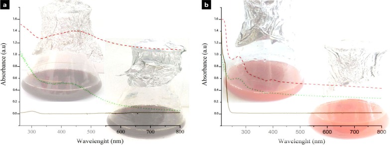
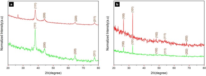
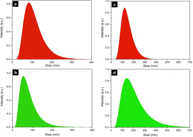
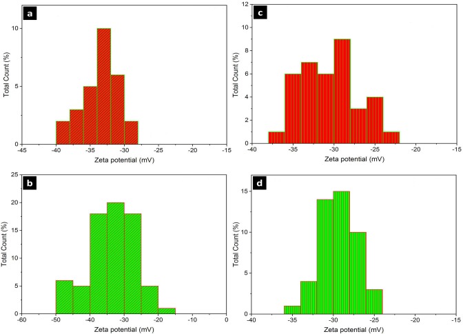
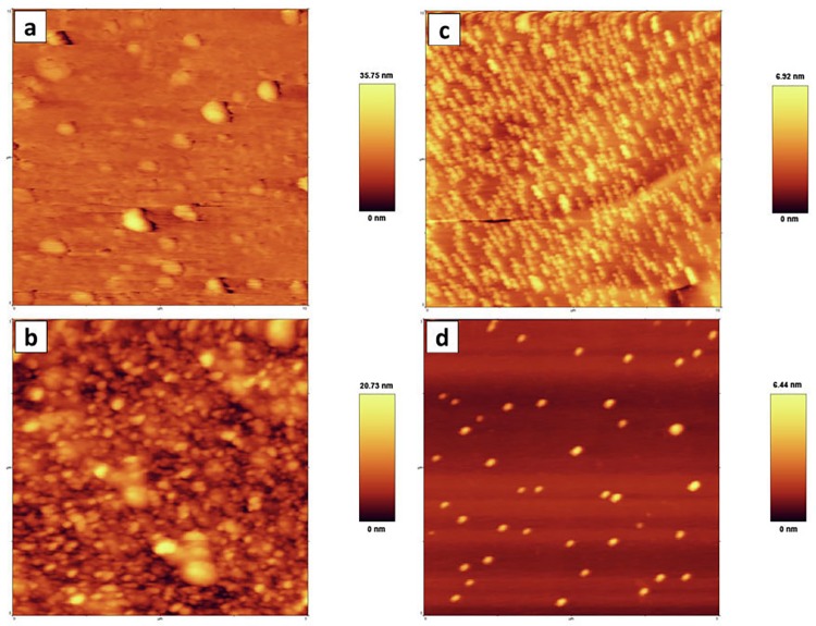
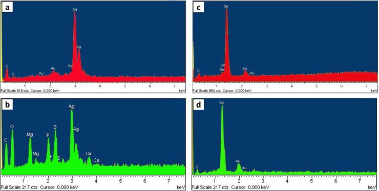
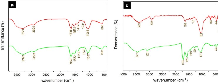
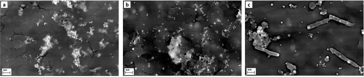
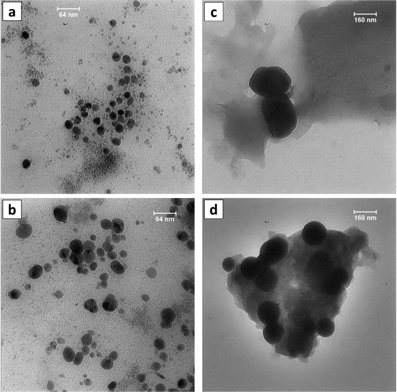
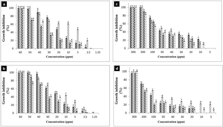
Halophiles are the organisms that thrive in extreme high salt environments. Despite the extensive studies on their biotechnological potentials, the ability of halophilic prokaryotes for the synthesis of nanoparticles has remained understudied. In this study, the archaeal and bacterial halophiles from a solar saltern were investigated for the intracellular/extracellular synthesis of silver and selenium nanoparticles. Silver nanoparticles were produced by the archaeal Haloferax sp. (AgNP-A, intracellular) and the bacterial Halomonas sp. (AgNP-B, extracellular), while the intracellular selenium nanoparticles were produced by the archaeal Halogeometricum sp. (SeNP-A) and the bacterial Bacillus sp. (SeNP-B). The nanoparticles were characterized by various techniques including UV-Vis spectroscopy, XRD, DLS, ICP-OES, Zeta potentials, FTIR, EDX, SEM, and TEM. The average particle size of AgNP-A and AgNP-B was 26.34 nm and 22 nm based on TEM analysis. Also, the characteristic Bragg peaks of face-centered cubic with crystallite domain sizes of 13.01 nm and 6.13 nm were observed in XRD analysis, respectively. Crystallographic characterization of SeNP-A and SeNP-B strains showed a hexagonal crystallite structure with domain sizes of 30.63 nm and 29.48 nm and average sizes of 111.6 nm and 141.6 nm according to TEM analysis, respectively. The polydispersity index of AgNP-A, AgNP-B, SeNP-A, and SeNP-B was determined as 0.26, 0.28, 0.27, and 0.36 and revealed high uniformity of the nanoparticles. All of the synthesized nanoparticles were stable and their zeta potentials were calculated as (mV): -33.12, -35.9, -31.2, and -29.34 for AgNP-A, AgNP-B, SeNP-A, and SeNP-B, respectively. The nanoparticles showed the antibacterial activity against various bacterial pathogens. The results of this study suggested that the (extremely) halophilic prokaryotes have great potentials for the green synthesis of nanoparticles.


Conflict of interest statement

The authors have declared that no competing interests exist.

Figures

Fig 1
Fig 1
UV-visible absorbance of the silver (a) and selenium (b) nanoparticles. Dash: archaea, dot: bacteria, solid: silver and selenium salts. Inlet: reduction of silver and selenium salts evidenced by color change of reaction mixture from to dark brown and red, respectively (up: archaea, down: bacteria).
Fig 2
Fig 2
X-ray diffraction (XRD) spectrum of the silver (a) and selenium (b) nanoparticles produced by archaeal (up) and bacterial (down) halophiles.
Fig 3
Fig 3. Dynamic light scattering (DLS) of silver and selenium nanoparticles.
a, AgNPs-A; b, AgNPs-B; c, SeNPs-A; d, SeNPs-B. AgNPs-A, silver nanoparticles synthesized by archaeal strain; AgNP-B, silver nanoparticles synthesized by bacterial strain; SeNP-A, selenium nanoparticles synthesized by archaeal strain; SeNP-B, selenium nanoparticles synthesized by bacterial strain.
Fig 4
Fig 4. Zeta potential analysis of the silver and selenium nanoparticles.
a, AgNPs-A; b, AgNPs-B; c, SeNPs-A; d, SeNPs-B. AgNPs-A, silver nanoparticles synthesized by archaeal strain; AgNP-B, silver nanoparticles synthesized by bacterial strain; SeNP-A, selenium nanoparticles synthesized by archaeal strain; SeNP-B, selenium nanoparticles synthesized by bacterial strain.
Fig 5
Fig 5. The AFM analysis of the silver and selenium nanoparticles.
a, AgNPs-A; b, AgNPs-B; c, SeNPs-A; d, SeNPs-B. AgNPs-A, silver nanoparticles synthesized by archaeal strain; AgNP-B, silver nanoparticles synthesized by bacterial strain; SeNP-A, selenium nanoparticles synthesized by archaeal strain; SeNP-B, selenium nanoparticles synthesized by bacterial strain.
Fig 6
Fig 6. Energy dispersive X-ray (EDX) spectrum of the silver and selenium nanoparticles.
a, AgNPs-A; b, AgNPs-B; c, SeNPs-A; d, SeNPs-B. AgNPs-A, silver nanoparticles synthesized by archaeal strain; AgNP-B, silver nanoparticles synthesized by bacterial strain; SeNP-A, selenium nanoparticles synthesized by archaeal strain; SeNP-B, selenium nanoparticles synthesized by bacterial strain.
Fig 7
Fig 7
FT-IR spectrum of the silver (a) and selenium (b) nanoparticels. up: AgNPs-A and SeNPs-A. Down: AgNP-B and SeNP-B. AgNPs-A, silver nanoparticles synthesized by archaeal strain; SeNP-A, selenium nanoparticles synthesized by archaeal strain; AgNP-B, silver nanoparticles synthesized by bacterial strain; SeNP-B, selenium nanoparticles synthesized by bacterial strain.
Fig 8
Fig 8
SEM images of the AgNP-A (a), SeNPs-A (b), and SeNPs-B (c) synthesized by the Haloferax, Halogeometricum and Bacillus strains, respectively.
Fig 9
Fig 9. TEM images of the silver and selenium nanoparticles.
a, AgNPs-A; b, AgNPs-B; c, SeNPs-A; d, SeNPs-B. AgNPs-A, silver nanoparticles synthesized by archaeal strain; AgNP-B, silver nanoparticles synthesized by bacterial strain; SeNP-A, selenium nanoparticles synthesized by archaeal strain; SeNP-B, selenium nanoparticles synthesized by bacterial strain.
Fig 10
Fig 10. Antibacterial activity of the silver and selenium nanoparticles.
a, AgNPs-A; b, AgNPs-B; c, SeNPs-A; d, SeNPs-B. AgNPs-A, silver nanoparticles synthesized by archaeal strain; AgNP-B, silver nanoparticles synthesized by bacterial strain; SeNP-A, selenium nanoparticles synthesized by archaeal strain; SeNP-B, selenium nanoparticles synthesized by bacterial strain. Microbial strains: sphere: E. coli; diamond: S. aureus; checker board: B. subtilis, dash: P. aeruginosa. Error bars indicate standard deviation for three replicates and different letters indicate significant difference between different concentrations (p<0.05).

References

    1. Oren A. Microbial life at high salt concentrations: phylogenetic and metabolic diversity. Saline Systems. 2008;15(4):2. - PMC - PubMed
    1. Ventosa A, Fernández AB, León MJ, Sánchez-Porro C, Rodriguez-Valera F. The Santa Pola saltern as a model for studying the microbiota of hypersaline environments. Extremophiles. 2014; 18(5), 811–24. 10.1007/s00792-014-0681-6 - DOI - PubMed
    1. Oren A. Industrial and environmental applications of halophilic microorganisms. Environmental technology. 2010; 31(8–9): 825–34. 10.1080/09593330903370026 - DOI - PubMed
    1. Jeevanandam J, Barhoum A, Chan YS, Dufresne A, Danquah MK. Review on nanoparticles and nanostructured materials: history, sources, toxicity and regulations. Beilstein journal of nanotechnology, 2018; 9(1): 1050–74. - PMC - PubMed
    1. Durán N, Marcato PD, Durán M, Yadav A, Gade A, Rai M. Mechanistic aspects in the biogenic synthesis of extracellular metal nanoparticles by peptide, bacteria, fungi, and plants. Applied Microbiology and Biotechnology. 2011; 90(5):1609–24 10.1007/s00253-011-3249-8 - DOI - PubMed

Publication types

Supplementary concepts

LinkOut - more resources