Repercussions of adjuvant-induced arthritis on body composition, soleus muscle, and heart muscle of rats
- PMID: 32130291
- PMCID: PMC7057929
- DOI: 10.1590/1414-431X20198969
Repercussions of adjuvant-induced arthritis on body composition, soleus muscle, and heart muscle of rats
Abstract
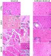
This study investigated the repercussions of adjuvant-induced arthritis (AIA) on body composition and the structural organization of the soleus and cardiac muscles, including their vascularization, at different times of disease manifestation. Male rats were submitted to AIA induction by intradermal administration of 100 μL of Mycobacterium tuberculosis (50 mg/mL), in the right hind paw. Animals submitted to AIA were studied 4 (AIA4), 15 (AIA15), and 40 (AIA40) days after AIA induction as well as a control group of animals not submitted to AIA. Unlike the control animals, AIA animals did not gain body mass throughout the evolution of the disease. AIA reduced food consumption, but only on the 40th day after induction. In the soleus muscle, AIA reduced the wet mass in a time-dependent manner but increased the capillary density by the 15th day and the fiber density by both 15 and 40 days after induction. The diameter of the soleus fiber decreased from the 4th day after AIA induction as well as the capillary/fiber ratio, which was most evident on the 40th day. Moreover, AIA induced slight histopathological changes in the cardiac muscle that were more evident on the 15th day after induction. In conclusion, AIA-induced changes in body composition as well as in the soleus muscle fibers and vasculature have early onset but are more evident by the 15th day after induction. Moreover, the heart may be a target organ of AIA, although less sensitive than skeletal muscles.
Figures


References
-
- Filippin LI, Vercelino R, Marroni NP, Xavier RM. Influência de processos redox na resposta inflamatória da artrite reumatóide. Rev Bras Reumatol. 2008;48:17–24. doi: 10.1590/S0482-50042008000100005. - DOI
-
- Vela P. Extra-articular manifestation of rheumatoid arthritis, Now. EMJ Rheumatol. 2014;1:103–12.
-
- Young A, Dixey J, Cox N, Davies P, Devlin J, Emery P, et al. How does functional disability in early rheumatoid arthritis (RA) affect patients and their lives? Results of 5 years of follow-up in 732 patients from the Early RA Study (ERAS) Rheumatology. 2000;39:603–611. doi: 10.1093/rheumatology/39.6.603. - DOI - PubMed
MeSH terms
LinkOut - more resources
Full Text Sources

