

Comparison of the Biograph Vision and Biograph mCT for quantitative 90Y PET/CT imaging for radioembolisation
- PMID: 32130554
- PMCID: PMC7056802
- DOI: 10.1186/s40658-020-0283-6
Comparison of the Biograph Vision and Biograph mCT for quantitative 90Y PET/CT imaging for radioembolisation
Abstract
Background: New digital PET scanners with improved time of flight timing and extended axial field of view such as the Siemens Biograph Vision have come on the market and are expected to replace current generation photomultiplier tube (PMT)-based systems such as the Siemens Biograph mCT. These replacements warrant a direct comparison between the systems, so that a smooth transition in clinical practice and research is guaranteed, especially when quantitative values are used for dosimetry-based treatment guidance. The new generation digital PET scanners offer increased sensitivity. This could particularly benefit 90Y imaging, which tends to be very noisy owing to the small positron branching ratio and high random fraction of 90Y. This study aims to determine the ideal reconstruction settings for the digital Vision for quantitative 90Y imaging and to evaluate the image quality and quantification of the digital Vision in comparison with its predecessor, the PMT-based mCT, for 90Y imaging in radioembolisation procedures.
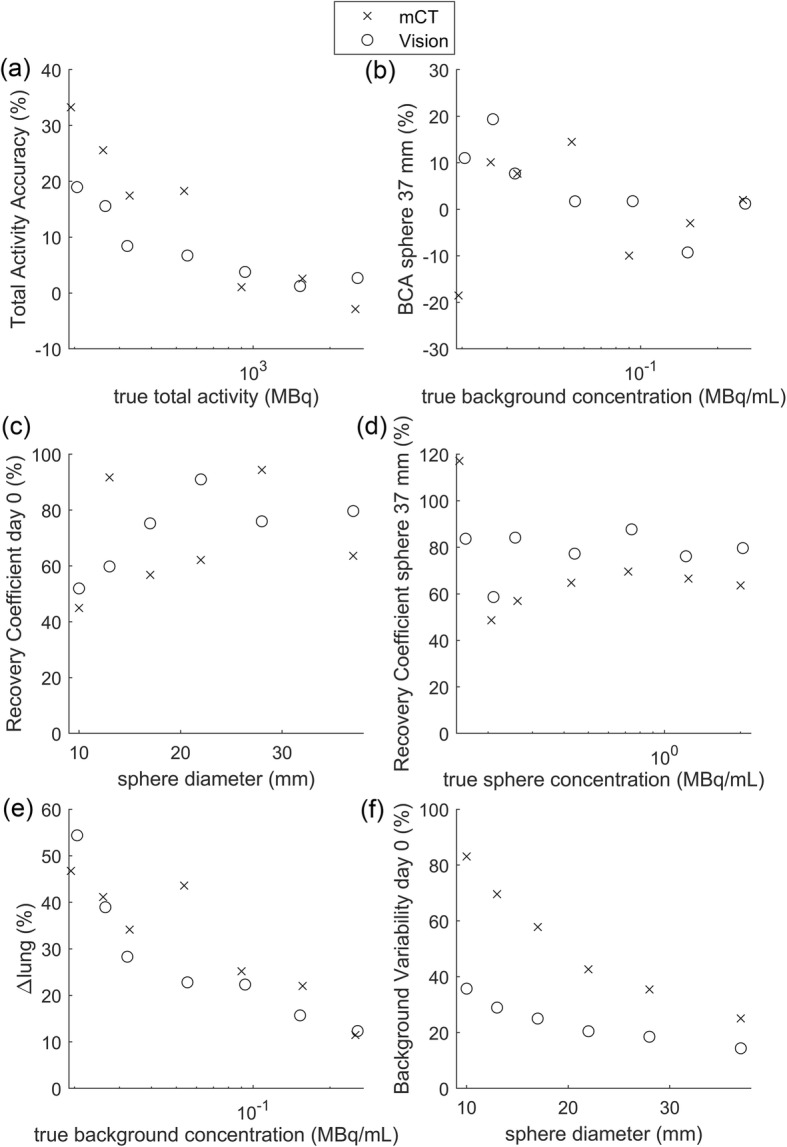
Methods: The NEMA image quality phantom was scanned to determine the ideal reconstruction settings for the Vision. In addition, an anthropomorphic phantom was scanned with both the Vision and the mCT, mimicking a radioembolisation patient with lung, liver, tumour, and extrahepatic deposition inserts. Image quantification of the anthropomorphic phantom was assessed by the lung shunt fraction, the tumour to non-tumour ratio, the parenchymal dose, and the contrast to noise ratio of extrahepatic depositions.
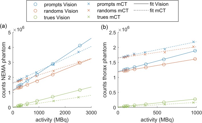
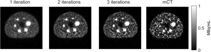
Results: For the Vision, a reconstruction with 3 iterations, 5 subsets, and no post-reconstruction filter is recommended for quantitative 90Y imaging, based on the convergence of the recovery coefficient. Comparing both systems showed that the noise level of the Vision is significantly lower than that of the mCT (background variability of 14% for the Vision and 25% for the mCT at 2.5·103 MBq for the 37 mm sphere size). For quantitative 90Y measures, such as needed in radioembolisation, both systems perform similarly.
Conclusions: We recommend to reconstruct 90Y images acquired on the Vision with 3 iterations, 5 subsets, and no post-reconstruction filter for quantitative imaging. The Vision provides a reduced noise level, but similar quantitative accuracy as compared with its predecessor the mCT.
Keywords: PET/CT; Radioembolisation; Yttrium-90.
Conflict of interest statement
ML is a consultant for BTG International and Terumo. The Department of Radiology and Nuclear Medicine of the UMC Utrecht receives research support and royalties from Quirem Medical.
Figures


References
-
- Elschot M, Vermolen BJ, Lam MGEH, de Keizer B, van den Bosch MAAJ, de Jong HWAM. Quantitative comparison of PET and Bremsstrahlung SPECT for imaging the in vivo yttrium-90 microsphere distribution after liver radioembolization. PLoS One. 2013;8(2):e55742. doi: 10.1371/journal.pone.0055742. - DOI - PMC - PubMed
-
- Alsultan AA, Smits MLJ, Barentsz MW, Braat AJAT, Lam MGEH. The value of yttrium-90 PET/CT after hepatic radioembolization: a pictorial essay. Clin Transl Imaging. 2019;7(4):303–312. doi: 10.1007/s40336-019-00335-2. - DOI
-
- Dewaraja Yuni K., Devasia Theresa, Kaza Ravi K., Mikell Justin K., Owen Dawn, Roberson Peter L., Schipper Matthew J. Prediction of Tumor Control in 90Y Radioembolization by Logit Models with PET/CT-Based Dose Metrics. Journal of Nuclear Medicine. 2019;61(1):104–111. doi: 10.2967/jnumed.119.226472. - DOI - PMC - PubMed
LinkOut - more resources
Full Text Sources
