

The value of navigation bronchoscopy in the diagnosis of peripheral pulmonary lesions: A meta-analysis
- PMID: 32130761
- PMCID: PMC7180606
- DOI: 10.1111/1759-7714.13373
The value of navigation bronchoscopy in the diagnosis of peripheral pulmonary lesions: A meta-analysis
Abstract
Background: To compare the diagnostic yield of peripheral pulmonary lesions (PPLs) with and without navigation system.
Methods: Studies dating from January 1990 to October 2019 were collected from databases. Diagnostic yield of navigation bronchoscopy and non-navigation bronchoscopy was extracted from comparative studies. Subgroup analysis was adopted to test diagnostic yield variation by lesion size, lobe location of the lesion, distance from the hilum, bronchus sign and nature of the lesion.
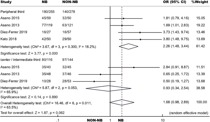
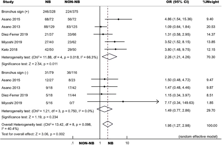
Results: In total, 2131 patients from 10 studies were enrolled into the study. Diagnostic yield of navigation bronchoscopy was statistically higher than non-navigation bronchoscopy for PPLs (odds ratio [OR] 1.69, 95% confidence interval [CI] 1.32, 2.18, P < 0.001), particularly for PPLs in the peripheral third lung (OR 2.26, 95% CI 1.48, 3.44, P < 0.001) and for bronchus sign positive PPLs (OR 2.26, 95% CI 1.21, 4.26, P = 0.011). Navigation bronchoscopy had better performance than non-navigation bronchoscopy when PPLs were ≤ 20 mm (OR 2.09, 95% CI 1.44, 3.03, P < 0.001). It also elevated diagnostic yield of malignant PPLs (OR 1.67, 95% CI 1.26, 2.22, P < 0.001) and PPLs in the bilateral upper lobes (OR 1.50, 95% CI 1.09, 2.08, P = 0.014).
Conclusions: Navigation bronchoscopy enhanced diagnostic yield when compared to non-navigation bronchoscopy, particularly for PPLs in the peripheral third lung, PPLs being bronchus sign positive, PPLs ≤ 20 mm, malignant PPLs and PPLs in the bilateral upper lobes.
Key points: The current study provided systematic evaluation on the diagnostic value of navigation bronchoscopy by comparing it with non-navigation bronchoscopy, and exploring the factors affecting the diagnostic yield.
Keywords: Diagnostic yield; electromagnetic navigation bronchoscopy (ENB); peripheral pulmonary lesions (PPLs); transbronchial lung biopsy (TBLB); virtual bronchoscopic navigation (VBN).
© 2020 The Authors. Thoracic Cancer published by China Lung Oncology Group and John Wiley & Sons Australia, Ltd.
Figures


 ) Included studies and (
 ) filled studies.


Similar articles
-
Virtual bronchoscopic navigation versus non-virtual bronchoscopic navigation assisted bronchoscopy for the diagnosis of peripheral pulmonary lesions: a systematic review and meta-analysis.Ther Adv Respir Dis. 2021 Jan-Dec;15:17534666211017048. doi: 10.1177/17534666211017048. Ther Adv Respir Dis. 2021. PMID: 34057861 Free PMC article.
-
Computed Tomography Bronchus Sign and the Diagnostic Yield of Guided Bronchoscopy for Peripheral Pulmonary Lesions. A Systematic Review and Meta-Analysis.Ann Am Thorac Soc. 2018 Aug;15(8):978-987. doi: 10.1513/AnnalsATS.201711-856OC. Ann Am Thorac Soc. 2018. PMID: 29877715
-
Electromagnetic navigation bronchoscopy versus virtual bronchoscopy navigation for improving the diagnosis of peripheral lung lesions: analysis of the predictors of successful diagnosis.Surg Today. 2022 Jun;52(6):923-930. doi: 10.1007/s00595-021-02398-z. Epub 2021 Oct 27. Surg Today. 2022. PMID: 34705111
-
Diagnosing a solitary pulmonary nodule using multiple bronchoscopic guided technologies: A prospective randomized study.Lung Cancer. 2019 Mar;129:48-54. doi: 10.1016/j.lungcan.2019.01.006. Epub 2019 Jan 16. Lung Cancer. 2019. PMID: 30797491 Clinical Trial.
-
Diagnostic yield of electromagnetic navigation bronchoscopy is highly dependent on the presence of a Bronchus sign on CT imaging: results from a prospective study.Chest. 2010 Dec;138(6):1316-21. doi: 10.1378/chest.09-2708. Epub 2010 Apr 30. Chest. 2010. PMID: 20435658
Cited by
-
The LungVision navigational platform for peripheral lung nodule biopsy and the added value of cryobiopsy.Thorac Cancer. 2021 Jul;12(13):2007-2012. doi: 10.1111/1759-7714.14003. Epub 2021 Jun 6. Thorac Cancer. 2021. PMID: 34096182 Free PMC article.
-
Advances in Diagnostic Bronchoscopy.Diagnostics (Basel). 2021 Oct 26;11(11):1984. doi: 10.3390/diagnostics11111984. Diagnostics (Basel). 2021. PMID: 34829331 Free PMC article. Review.
-
Novel diagnostic processes and challenges in bronchoscopy.Pathol Oncol Res. 2024 May 21;30:1611774. doi: 10.3389/pore.2024.1611774. eCollection 2024. Pathol Oncol Res. 2024. PMID: 38835723 Free PMC article. Review.
-
4D Electromagnetic Navigation Bronchoscopy for the Sampling of Pulmonary Lesions: First European Real-Life Experience.Lung. 2021 Oct;199(5):493-500. doi: 10.1007/s00408-021-00477-z. Epub 2021 Sep 25. Lung. 2021. PMID: 34562105 Free PMC article.
-
Ultrathin bronchoscope combined with virtual bronchoscopic navigation and endobronchial ultrasound for the diagnosis of peripheral pulmonary lesions with or without fluoroscopy: A randomized trial.Thorac Cancer. 2021 Jun;12(12):1864-1872. doi: 10.1111/1759-7714.13995. Epub 2021 May 6. Thorac Cancer. 2021. PMID: 33956409 Free PMC article. Clinical Trial.
References
-
- Rivera MP, Mehta AC, Wahidi MM. Establishing the diagnosis of lung cancer: Diagnosis and management of lung cancer, 3rd ed: American College of Chest Physicians evidence‐based clinical practice guidelines. Chest 2013; 143: e142S–65S. - PubMed
-
- Bai C, Choi CM, Chu CM et al Evaluation of pulmonary nodules: Clinical practice consensus guidelines for Asia. Chest 2016; 150: 877–93. - PubMed
-
- Schreiber G, McCrory DC. Performance characteristics of different modalities for diagnosis of suspected lung cancer: Summary of published evidence. Chest 2003; 123: 115S–28S. - PubMed
Publication types
MeSH terms
LinkOut - more resources
Full Text Sources
Medical
