. 2020 Apr;24(7):4312-4323.
doi: 10.1111/jcmm.15091. Epub 2020 Mar 4.

iNOS-inhibitor driven neuroprotection in a porcine retina organ culture model

Affiliations

iNOS-inhibitor driven neuroprotection in a porcine retina organ culture model

José Hurst et al. J Cell Mol Med. 2020 Apr.

Abstract

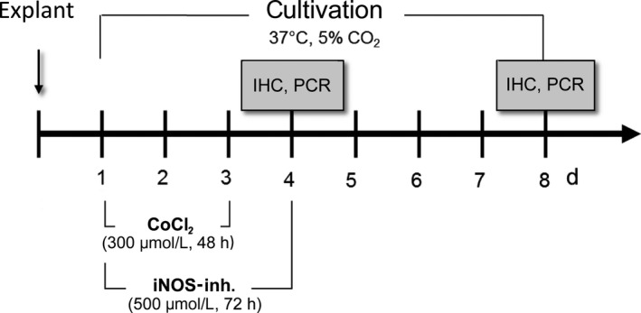
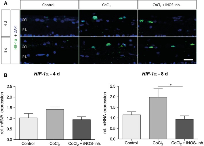
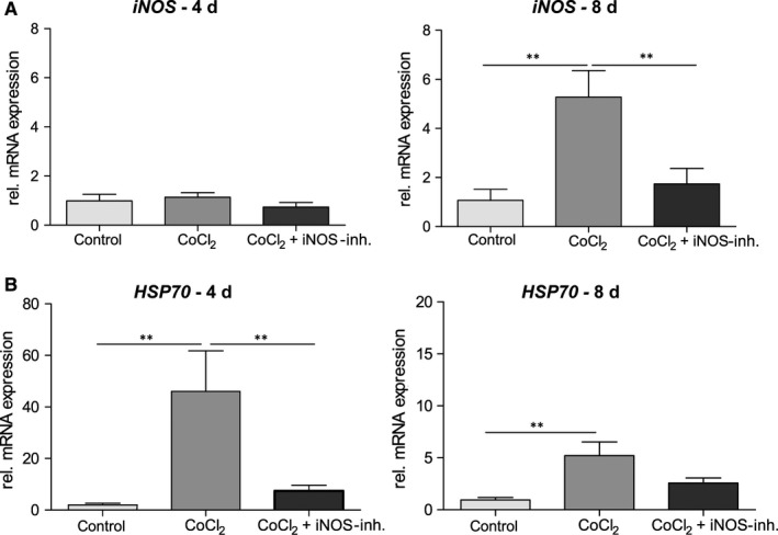
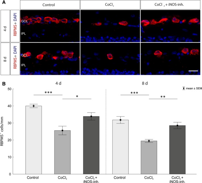
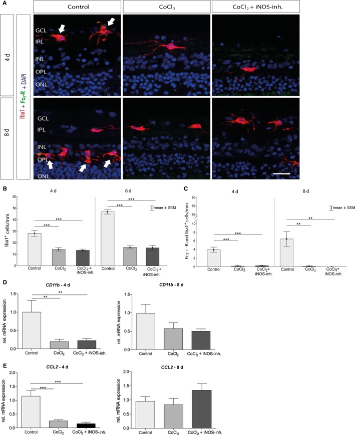
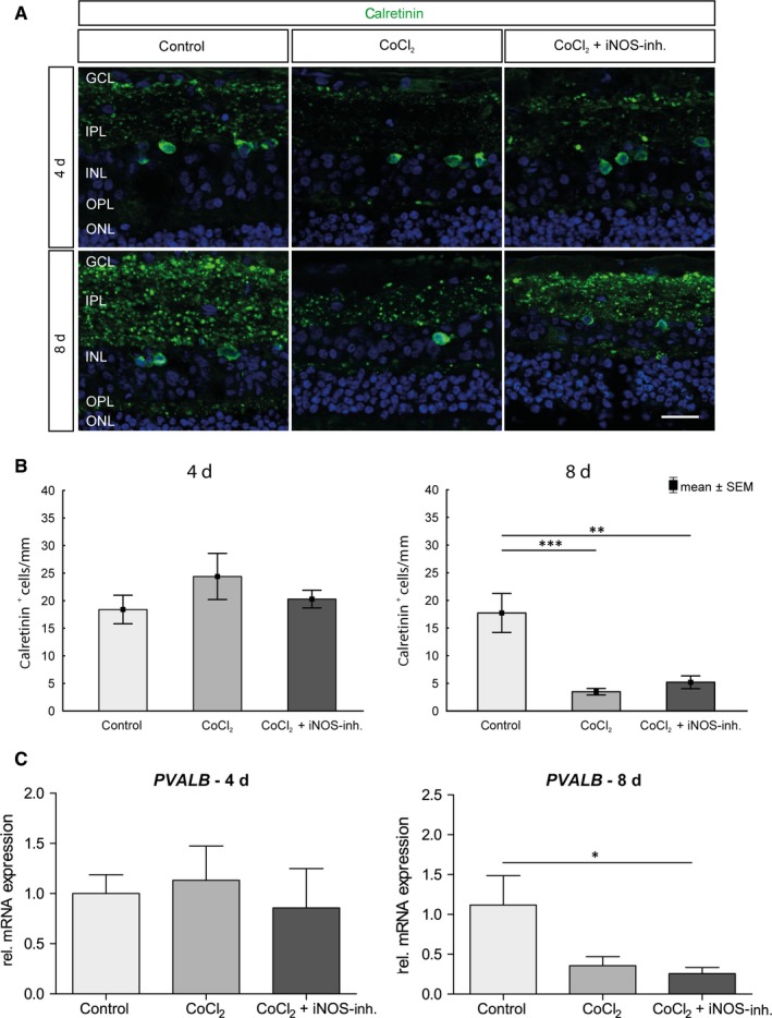
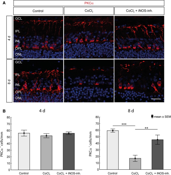
Nitrite oxide plays an important role in the pathogenesis of various retinal diseases, especially when hypoxic processes are involved. This degeneration can be simulated by incubating porcine retinal explants with CoCl2 . Here, the therapeutic potential of iNOS-inhibitor 1400W was evaluated. Degeneration through CoCl2 and treatment with the 1400W were applied simultaneously to porcine retinae explants. Three groups were compared: control, CoCl2 , and CoCl2 + iNOS-inhibitor (1400W). At days 4 and 8, retinal ganglion cells (RGCs), bipolar, and amacrine cells were analysed. Furthermore, the influence on the glia cells and different stress markers were evaluated. Treatment with CoCl2 resulted in a significant loss of RGCs already after 4 days, which was counteracted by the iNOS-inhibitor. Expression of HIF-1α and its downstream targets confirmed the effective treatment with 1400W. After 8 days, the CoCl2 group displayed a significant loss in amacrine cells and also a drastic reduction in bipolar cells was observed, which was prevented by 1400W. The decrease in microglia could not be prevented by the inhibitor. CoCl2 induces strong degeneration in porcine retinae by mimicking hypoxia, damaging certain retinal cell types. Treatment with the iNOS-inhibitor counteracted these effects to some extent, by preventing loss of retinal ganglion and bipolar cells. Hence, this inhibitor seems to be a very promising treatment for retinal diseases.

Keywords: hypoxia; iNOS-inhibitor 1400W; organ culture; retina; retinal ganglion cells.


Conflict of interest statement

The authors confirm that there are no conflicts of interest.

Figures

Figure 1
Figure 1
Procedure of the 1400W treatment in the CoCl2 degeneration model. CoCl2, cobalt chloride; IHC, immunohistochemistry; iNOS‐inh., iNOS‐inhibitor 1400W
Figure 2
Figure 2
iNOS‐inhibitor mediated effect on hypoxia‐induced HIF‐1α expression. A, Representative pictures of HIF‐1α staining in the ganglion cell layer. HIF‐1α+ cells are marked in green and cell nuclei in blue. The addition of CoCl2 induced an increase or stabilization of the alpha subunit of the transcription factor. Under treatment with the iNOS inhibitor, the HIF‐1a levels were not lower compared with the CoCl2 group after 4 d. In addition, at 8 d of cultivation, comparable amounts of HIF‐1α were detected in CoCl2 and in the 1400W‐treated group. B, After 4 d, no significant differences in HIF‐1α mRNA expression were observed. At 8 d, the relative mRNA expression by CoCl2 was twofold increased and could be lowered to the initial level by treatment with 1400W. Abbreviations: GCL, ganglion cell layer; IPL, inner plexiform layer. Scale bar = 20 µm. All data are shown as mean ± SEM; *P < .05
Figure 3
Figure 3
mRNA expression of the HIF‐1α target genes. A, CoCl2 had a significant effect on iNOS mRNA expression after 4 d and the additional treatment with 1400W only lead to a small decrease in mRNA. After 8 d, CoCl2 induced a fourfold increase in iNOS mRNA expression, this could be prevented by the 1400W treatment. B, After 4 d, a massive increase in mRNA expression of HSP70 was observed in the CoCl2 retinas, which was significantly reduced by treatment with 1400W. After 8 d, a significantly increased expression was still observed, but not as pronounced as at the previous time. However, the protective effect of the inhibitor was still detectable after 8 d. All data are shown as mean ± SEM; **P < .01
Figure 4
Figure 4
Rescue of retinal ganglion cells after CoCl2‐induced degeneration. A, Representative images of the immunohistological staining. RGCs were stained with an antibody against RBPMS (red) and cell nuclei with DAPI (blue). A significant loss of RGCs in the untreated degeneration groups (CoCl2) was observed over the cultivation period of 4 and 8 d. B, After 4 d, neuroprotection of the RGCs was observed by treatment with the iNOS‐inhibitor compared with the CoCl2 group. Even after 8 d of cultivation, a protection of the RGCs by 1400W could be noticed. The retinae of the treatment groups contained significantly more RGCs than the untreated retinae. GCL, ganglion cell layer; IPL, inner plexiform layer. Scale bar = 20 µm. All data are shown as mean ± SEM; *P < .05; **P < .01; ***P < .001
Figure 5
Figure 5
CoCl2 degeneration irreversibly reduces the microglia. A, Microglia were stained with anti‐Iba1 (red) on day 4 and 8 of cultivation. Fcy‐R (green, arrows) in combination with Iba1 served as an activity marker of the microglia. Cell nuclei are shown in blue. B, The addition of CoCl2 triggered a significant loss of microglia in comparison with controls at both times. In the 1400W‐treated group also, significantly fewer microglia were present than in the control. C, In addition, the number of activated microglia was significantly lower in the CoCl2 group than in the control group after 4 and 8 d. Again, treatment with the iNOS‐inhibitor had no protective effect. D, The relative CD11b mRNA expression was also significantly reduced in the CoCl2 group, after 4 and 8 d of cultivation. The 1400W treatment did not result in an improvement compared with control at both times. E, The analysis of the relative CCL2 mRNA expression showed that it was significantly reduced after 4 d in retinae of the CoCl2 group. Again, mRNA expression could not be altered by the 1400W. After 8 d of cultivation, no differences between the groups were observed. GCL, ganglion cell layer; INL, inner nuclear layer; IPL, inner plexiform layer; ONL, outer nuclear layer; OPL, outer plexiform layer. Scale bar = 20 µm. All data are shown as mean ± SEM; P</i> < .01; "?>**P < .01; ***P < .001
Figure 6
Figure 6
No positive influence of 1400W treatment on amacrine cells. A, Representative pictures of amacrine cell population in retinal explants. Amacrine cells were labelled with antibodies against calretinin (green). Nuclei were stained with DAPI (blue). B, On day 4, no significant differences could be observed between the groups. On day 8, a significant loss of amacrine cells in retinae of the CoCl2 group was detected. The iNOS inhibitor treatment could not protect the amacrine cells. C, No differences in the relative expression of PVALB mRNA were observed on day 4. On day 8, the relative expression of PVALB was reduced in all groups compared with the control and significantly in comparison with the iNOS‐inh. treatment. GCL, ganglion cell layer; INL, inner nuclear layer; IPL, inner plexiform layer; ONL, outer nuclear layer; OPL, outer plexiform layer. Scale bar = 20 µm. All data are shown as mean ± SEM. *P < .05; **P < .01; ***P < .001
Figure 7
Figure 7
Protection of bipolar cells against CoCl2‐induced degeneration. A, Nuclei were visualized with DAPI staining (blue), and bipolar cells were stained with an antibody against PKCα (red). B, No significant loss of bipolar cells in the untreated degeneration groups could be detected during the cultivation period of 4 d. After 8 d, a significant loss was observed. However, protection of the bipolar cells by 1400W was observed. The retinae of the treatment group contained significantly more bipolar cells than the untreated retinae. GCL, ganglion cell layer; INL, inner nuclear layer; IPL, inner plexiform layer; ONL, outer nuclear layer; OPL, outer plexiform layer. Scale bar = 20 µm. All data are shown as mean ± SEM; **P < .01; ***P < .001

Similar articles

Cited by

References

    1. Daneshian M, Busquet F, Hartung T, Leist M. Animal use for science in Europe. Altex‐Altern Anim Ex. 2015;32:261‐274. - PubMed
    1. Kuehn S, Hurst J, Jashari A, et al. A novel NMDA triggered porcine organ culture induces retinal ganglion cell apoptosis – chances for replacement of animal experiments. Altern Lab Anim. 2016;44:557‐568. - PubMed
    1. Kuehn S, Hurst J, Rensinghoff F, et al. Degenerative effects of cobalt‐chloride treatment on neurons and microglia in a porcine retina organ culture model. Exp Eye Res. 2017;155:107‐120. - PubMed
    1. Maliha AM, Kuehn S, Hurst J, et al. Diminished apoptosis in hypoxic porcine retina explant cultures through hypothermia. Sci Rep. 2019;9(1):4898. - PMC - PubMed
    1. Hurst J, Kuehn S, Jashari A, et al. A novel porcine ex vivo retina culture model for oxidative stress induced by H2O2 . Altern Lab Anim. 2017;45:11‐25. - PubMed

Publication types

MeSH terms

Associated data