. 2020 May 21;27(5):525-537.e6.
doi: 10.1016/j.chembiol.2020.02.003. Epub 2020 Mar 3.

Targeting the PI5P4K Lipid Kinase Family in Cancer Using Covalent Inhibitors

Affiliations

Targeting the PI5P4K Lipid Kinase Family in Cancer Using Covalent Inhibitors

Sindhu Carmen Sivakumaren et al. Cell Chem Biol. .

Abstract

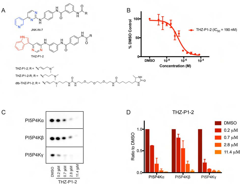
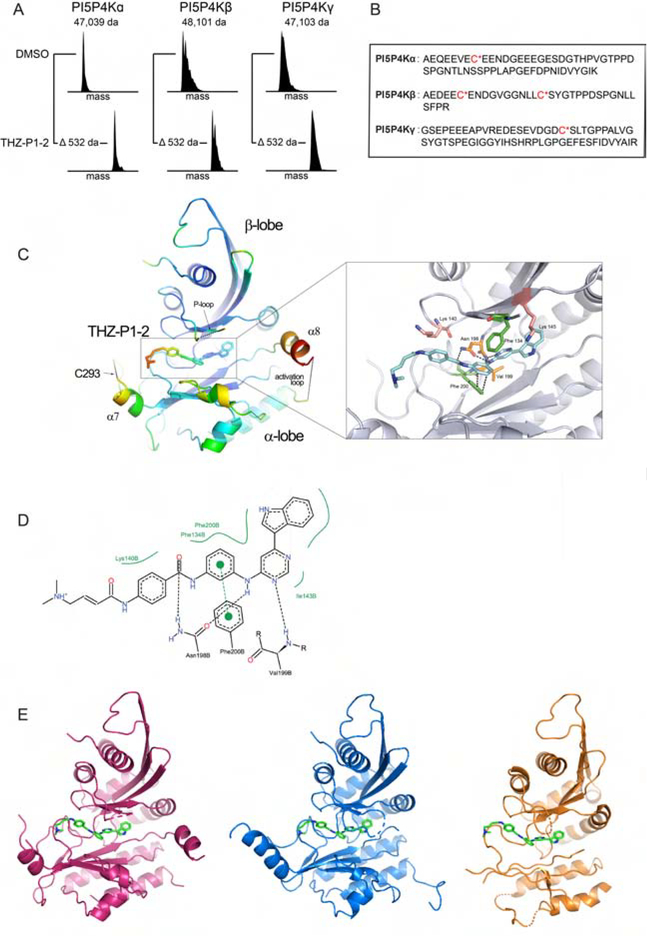
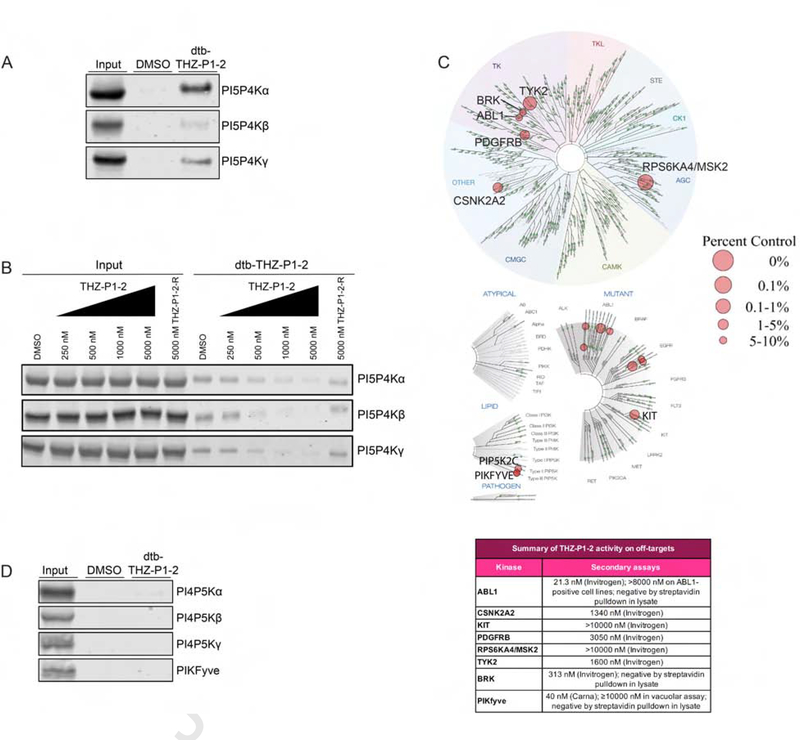
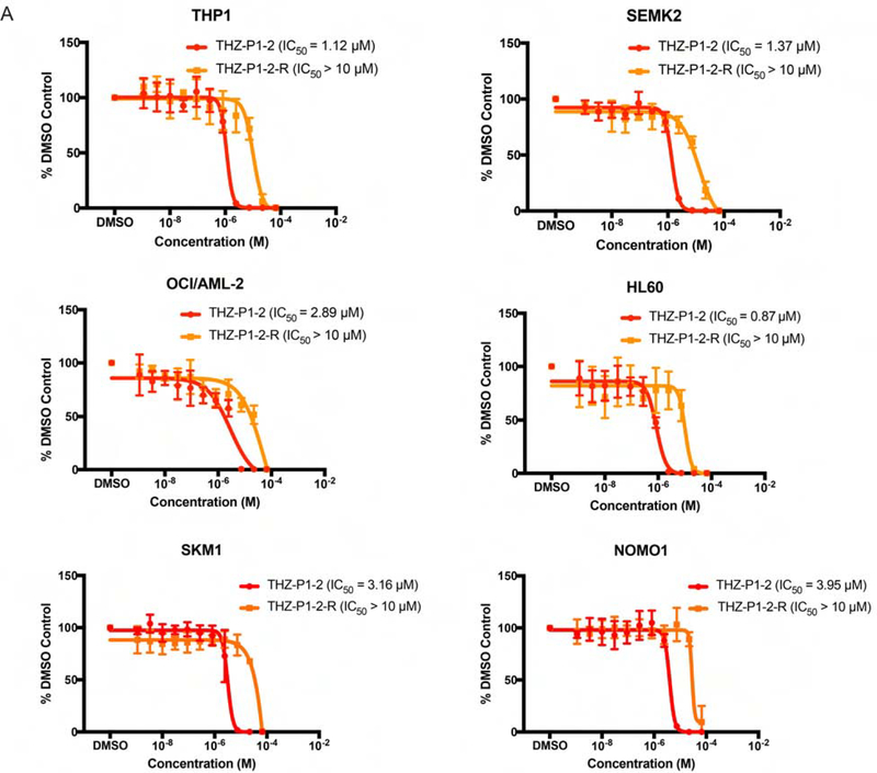
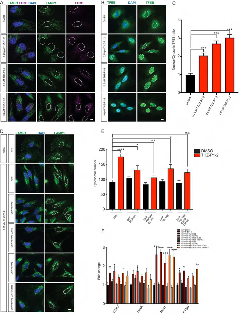
The PI5P4Ks have been demonstrated to be important for cancer cell proliferation and other diseases. However, the therapeutic potential of targeting these kinases is understudied due to a lack of potent, specific small molecules available. Here, we present the discovery and characterization of a pan-PI5P4K inhibitor, THZ-P1-2, that covalently targets cysteines on a disordered loop in PI5P4Kα/β/γ. THZ-P1-2 demonstrates cellular on-target engagement with limited off-targets across the kinome. AML/ALL cell lines were sensitive to THZ-P1-2, consistent with PI5P4K's reported role in leukemogenesis. THZ-P1-2 causes autophagosome clearance defects and upregulation in TFEB nuclear localization and target genes, disrupting autophagy in a covalent-dependent manner and phenocopying the effects of PI5P4K genetic deletion. Our studies demonstrate that PI5P4Ks are tractable targets, with THZ-P1-2 as a useful tool to further interrogate the therapeutic potential of PI5P4K inhibition and inform drug discovery campaigns for these lipid kinases in cancer metabolism and other autophagy-dependent disorders.

Keywords: PI5P4K; autophagy; cancer; covalent inhibitor; drug discovery; kinase; phosphoinositide.


Conflict of interest statement

Declaration of Interests J.A.M. is a member of the scientific advisory board (SAB) of 908 Devices. L.C.C. is a founder and member of the Board of Directors of Agios Pharmaceuticals and is a founder and receives research support from Petra Pharmaceuticals. These companies are developing novel therapies for cancer. N.S.G. is a founder, SAB member, and equity holder in Gatekeeper, Syros, Petra, C4, B2S, and Soltego. The Gray lab receives or has received research funding from Novartis, Takeda, Astellas, Taiho, Janssen, Kinogen, Voronoi, Her2llc, Deerfield, and Sanofi. N.S.G., T.Z., and N.P.K. are inventors on a patent application covering chemical matter in this publication owned by the Dana-Farber Cancer Institute.

Figures

Figure 1.
Figure 1.. Chemoproteomic profiling and synthetic chemistry approaches reveal PI5P4K inhibitor THZ-P1-2.
(A) Chemical structures of compounds JNK-IN-7 as previously reported in Zhang et al. (2012) and THZ-P1-2 with its reversible and desthiobiotinylated counterparts. See also Fig. S1. (B) THZ-P1-2 potently inhibits PI5P4Kα kinase activity in the ADP-Glo luminescence assay. Data shown are the mean of two biological replicates ± SEM. (C) THZ-P1-2 shows potent inhibition of kinase activity of all three PI5P4K isoforms in a radiometric TLC assay measuring radiolabeled PI-4,5-P2. TLC image shown is representative of two independent experiments. (D) Quantification of (C). Radiolabeled PI-4,5-P2 spots were imaged by autoradiography and quantified by densitometry.
Figure 2.
Figure 2.. THZ-P1-2 binds covalently to all isoforms of the PI5P4K family on unique cysteine residues located on a disordered loop outside the kinase domain.
(A) Electrospray mass spectrometry of recombinant PI5P4Kα/β/γ incubated with THZ-P1-2 demonstrates covalent labeling of PI5P4K isoforms. See also Fig. S2B. (B) Subsequent protease digestion and tandem mass spectrometry confirms that THZ-P1-2 covalently labels cysteine residues. See also Fig. S2A. (C) Crystal structure of PI5P4Kα in complex with THZ-P1-2, colored according to B factor and shown with covalent warhead extended out towards the covalently-targeted cysteine, C293 (labeled; not resolved in crystal structure). See also Table S1. (D) Ligand interaction map of THZ-P1-2 with residues in the ATP-binding pocket of PI5P4Kα. Related to Fig. S2C–D. (E) Modeled THZ-P1-2 binding showed for all three isoforms based on alignment of published PI5P4Kβ (1BO1; Rao et al., 1998) and γ (2GK9; Uppenberg et al., 2006) structures with obtained PI5P4Kα structure. PI5P4Kα (magenta), PI5P4Kβ (blue), PI5P4Kγ (orange).
Figure 3.
Figure 3.. Cellular on-target engagement and selectivity profile of THZ-P1-2.
(A) A desthiobiotinylated analog of THZ-P1-2 irreversibly engages all PI5P4K isoforms in a streptavidin pulldown in HEK293T lysate. (B) THZ-P1-2, but not its reversible analog, exhibits dose-dependent on-target engagement of PI5P4K isoforms in a competitive streptavidin pulldown with 1 μM desthiobiotinylated-THZ-P1-2. See also Fig. S3B. (C) Kinome selectivity profile of THZ-P1-2. Complete dataset for off-targets included as Table S2. See also Fig. S4A–C. (D) THZ-P1-2 does not engage the PI4P5Ks or PIKfyve in a streptavidin pulldown. See also Fig. S3C.
Figure 4.
Figure 4.. Preliminary cancer cell line profiling with THZ-P1-2 to identify potential PI5P4K dependencies.
(A) A panel of AML and ALL cell lines were treated with THZ-P1-2 and THZ-P1-2-R for 72h and cell proliferation was measured with a Cell-Titer Glo luminescence assay. Data shown are the mean of three biological replicates ± SEM.
Figure 5.
Figure 5.. Inhibition of PI5P4K with THZ-P1-2 leads to lysosomal-autophagosomal defects and increased TFEB activation
(A) Similar to genetic loss of PIP4K2A/B, inhibition of PI5P4K activity with THZ-P1-2 increases LAMP1-positive lysosomal size, number and contact with LC3B-positive autophagosomes. HeLa cells were cultured overnight with either DMSO, 0.25 μM, 0.5 μM or 1.0 μM THZ-P1-2 and stained for LC3B (magenta) or LAMP1 (green) with nuclei in blue. Scale bars, 10 μm. See also Fig. S5. (B) Inhibition of PI5P4K activity with THZ-P1-2 increases TFEB nuclear localization. HeLa cells were cultured overnight with either DMSO, 0.25 μM, 0.5 μM or 1.0 μM THZ-P1-2 and stained for TFEB (green) with nuclei in blue. Scale bars, 10 μm. (C) Quantification of results in (B). The intensity of TFEB immunofluorescence was quantified in the nucleus and the cytoplasm, and used to calculate the ratio. Statistical significance determined by ANOVA (***p < 0.0005) with Dunnett multiple comparison post-test. Each group was compared to control control HeLa cells treated with DMSO, (n ≥ 30). (D) Expression of PI5P4K cysteine to serine mutants alleviates lysosomal dysfunction induced by THZ-P1-2 treatment. HeLa cells were infected with viruses expressing GFP, GFP-PI5P4Kα, GFP- PI5P4Kα C293S, GFP-PI5P4Kβ, or GFP-PI5P4Kβ C307S C318S and treated with either DMSO or 250 nM THZ-P1-2 overnight. Expression of both the PI5P4Kα and PI5P4Kβ cysteine to serine mutants alleviated dysfunctional lysosomes (green) with nuclei in blue. Scale bars, 10 μm. (E) Quantification of results in (D). The number of lysosomes was quantified per cell. Statistical significance determined by ANOVA (***p < 0.0005) with Dunnett multiple comparison post-test. Each group was compared to control HeLa cells expressing GFP and treated with DMSO, (n ≥ 30). (F) HeLa cells expressing GFP, GFP-PI5P4Kα, GFP- PI5P4Kα C293S, GFP-PI5P4Kβ, or GFP-PI5P4Kβ C307S C318S were treated with either DMSO or 250 nM THZ-P1-2 overnight for 16 hours and subsequently harvested for qPCR of TFEB targets. Fold change is calculated by comparison to HeLa cells expressing GFP treated with DMSO. *p < 0.05, Student’s t-test, (n ≥ 8).

Similar articles

Cited by

References

    1. Adams PD, Afonine PV, Bunkóczi G, Chen VB, Davis IW, Echols N, … Zwart PH (2010). PHENIX: A comprehensive Python-based system for macromolecular structure solution. Acta Crystallographica Section D: Biological Crystallography, 66(2), 213–221. 10.1107/S0907444909052925 - DOI - PMC - PubMed
    1. Al-ramahi I, Srinivas S, Giridharan P, Chen Y, Patnaik S, Safren N, … Marugan JJ (2017). Inhibition of PIP4K g ameliorates the pathological effects of mutant huntingtin protein, 1–25. - PMC - PubMed
    1. Balla T, Szentpetery Z, & Kim YJ (2009). Phosphoinositide Signaling: New Tools and Insights. Physiology, 24(4), 231–244. 10.1152/physiol.00014.2009 - DOI - PMC - PubMed
    1. Braekeleer ED, Douet-Guilbert N, Rowe D, Bown N, Morel F, Berthou C, … Braekeleer MD (2011). ABL1 fusion genes in hematological malignancies: A review. European Journal of Haematology,86(5), 361–371. doi: 10.1111/j.1600-0609.2011.01586.x - DOI - PubMed
    1. Browne CM, Jiang B, Ficarro SB, Doctor ZM, Johnson JL, Card JD, … Marto JA (2018). A Chemoproteomic Strategy for Direct and Proteome-Wide Covalent Inhibitor Target-Site Identification. Journal of the American Chemical Society,141(1), 191–203. doi:10.1021/jacs.8b07911 - DOI - PMC - PubMed

Publication types

MeSH terms

Substances

LinkOut - more resources