

doi: 10.1016/j.antiviral.2020.104759.
Epub 2020 Mar 1.
A potential role for integrins in host cell entry by SARS-CoV-2
Affiliations
- PMID: 32130973
- PMCID: PMC7114098
- DOI: 10.1016/j.antiviral.2020.104759
Item in Clipboard
A potential role for integrins in host cell entry by SARS-CoV-2
Antiviral Res.
2020 May.
Abstract
- •
Integrin may act as an alternative receptor for SARS-CoV-2 and could be implicated in its transmission and pathology.
- •
The spike protein of SARS-CoV-2 acquired a RGD motif known to bind integrins. This motif is absent from other coronaviruses.
- •
The integrin-binding motif is present at the surface of the spike protein, close to the ACE2 receptor-binding region.
- •
Integrin binding may be a promising therapeutics target, and should be tested experimentally.
Figures
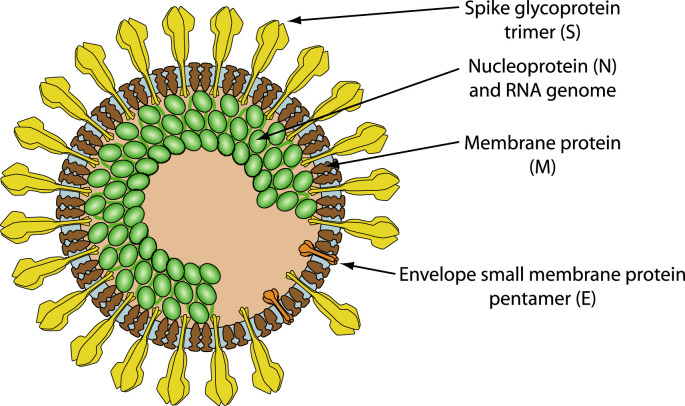
Schematic representation of SARS coronavirus virion (Hulo et al., 2011), based on cryo-electron microscopy (Neuman et al., 2011).
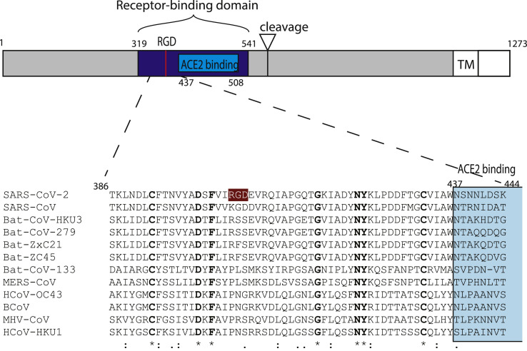
Schematic representation of SARS-CoV-2 S-protein with a focus on the receptor-binding domain. The sequences of 12 betacoronavirus were aligned using MAFFT (Katoh et al., 2019). The receptor-binding domain and the ACE2 receptor-binding region are colored in blue and light blue, respectively. The RGD motif of SARS-CoV-2 is highlighted in color. Numbers refer to the SARS-CoV-2 spike protein sequence.
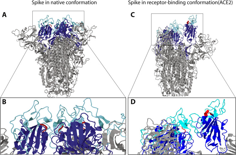
A) and B) Model of SARS-CoV-2 structure provided by SWISSMODEL and visualized with Jmol. A) The mushroom-like fold of the model is the classical one in absence of ligand binding. Ligand binding causes a drastic conformational change leading to the protrusion of one of the trimeric binding domains, further exposing the RGD-loop. The receptor-binding domain has been colored in blue, with a focus in light blue on the part binding the receptor ACE2. The RGD motif is colored in red. B) Enlarged view of the receptor-binding domain. The region demonstrated to bind ACE2, as well as the RGD-loops, are located at the surface of the domain even in the absence of ligand. Same colors as in A. C) and D) Model of SARS-CoV-2 structure in the conformational state of ACE2-binding provided by SWISSMODEL and visualized with Jmol. C) The receptor-binding domain of the trimer is in the “up” conformation exposing the RGD motif. Same color as in Fig. 2; ACE2 is not represented. D) Close-up view of the receptor-binding domain, highlighting the location of the RGD motif at the very surface of the domain. Same colors as in A.
References
-
- Betacoronavirus ViralZone page. https://viralzone.expasy.org/764?outline=all_by_species [WWW document], n.d. accessed 2.20.20.
Publication types
MeSH terms
Substances
Grants and funding
LinkOut - more resources
Full Text Sources
Other Literature Sources
Miscellaneous
