. 2020 Mar 2;12(3):576.
doi: 10.3390/cancers12030576.

NUDT7 Loss Promotes KrasG12D CRC Development

Affiliations

NUDT7 Loss Promotes KrasG12D CRC Development

Jinsoo Song et al. Cancers (Basel). .

Abstract

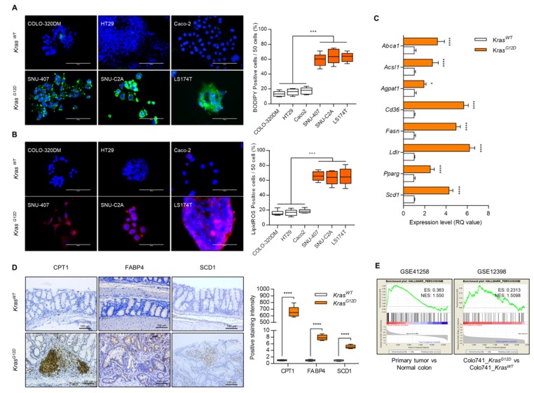
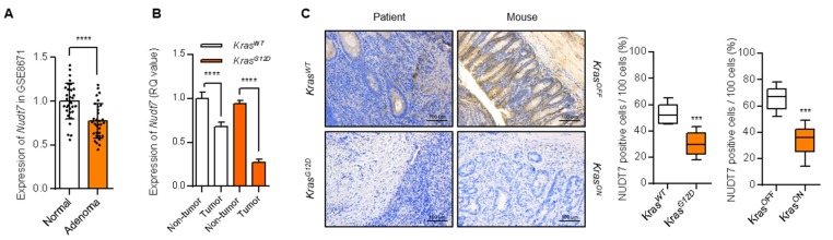
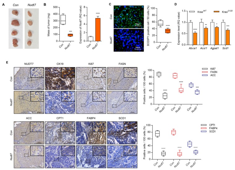
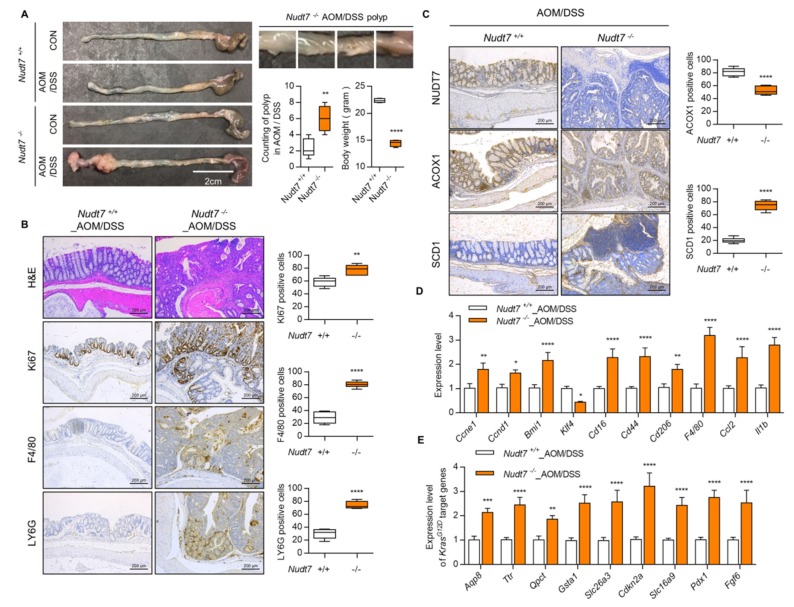
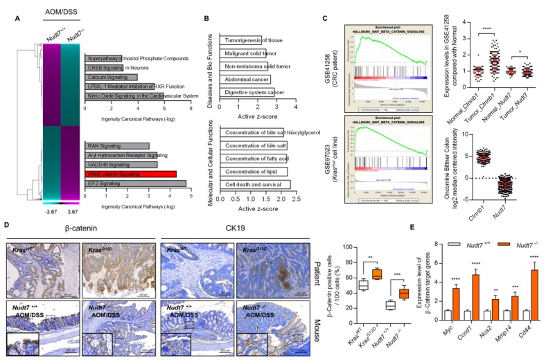
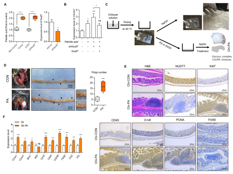
Studies have suggested that dysregulation of peroxisomal lipid metabolism might play an important role in colorectal cancer (CRC) development. Here, we found that KrasG12D-driven CRC tumors demonstrate dysfunctional peroxisomal b-oxidation and identified Nudt7 (peroxisomal coenzyme A diphosphatase NUDT7) as one of responsible peroxisomal genes. In KrasG12D-driven CRC tumors, the expression level of Nudt7 was significantly decreased. Treatment of azoxymethane/dextran sulfate sodium (AOM/DSS) into Nudt7 knockout (Nudt7-/-) mice significantly induced lipid accumulation and the expression levels of CRC-related genes whereas xenografting of Nudt7-overexpressed LS-174T cells into mice significantly reduced lipid accumulation and the expression levels of CRC-related genes. Ingenuity pathway analysis of microarray using the colon of Nudt7-/- and Nudt7+/+ mice treated with AOM/DSS suggested Wnt signaling as one of activated signaling pathways in Nudt7-/- colons. Upregulated levels of β-catenin were observed in the colons of KrasG12D and AOM/DSS-treated Nudt7-/- mice and downstream targets of β-catenin such as Myc, Ccdn1, and Nos2, were also significantly increased in the colon of Nudt7-/- mice. We observed an increased level of palmitic acid in the colon of Nudt7-/- mice and attachment of palmitic acid-conjugated chitosan patch into the colon of mice induced the expression levels of b-catenin and CRC-related genes. Overall, our data reveal a novel role for peroxisomal NUDT7 in KrasG12D-driven CRC development.

Keywords: colorectal cancer; palmitic acid; peroxisomal coenzyme A diphosphatase NUDT7 (NUDT7); peroxisome; β-catenin.


Conflict of interest statement

The authors declare no potential conflicts of interest.

Figures

Figure 1
Figure 1
Dysfunction of lipid metabolism in KrasG12D colorectal cancer (CRC) cells. (A) BODIPY staining and (B) lipid reactive oxygen species (ROS) staining using KrasG12D and KrasWT cell lines. Positive cells were counted for every 50 cells in 3 different fields at 400× magnification. Results shown are representative of at least 3 independent experiments. Scale bars: 100 μm. (C) Expression level of genes involved in lipid metabolism in KrasG12D CRC cells and presented as the fold change of KrasWT CRC cells. Rn18s was used as an endogenous control. Results are representative of at least 3 independent experiments. (D) Immunohistochemical staining with CPT1, FABP4, and SCD1, and positive cells were counted (n = 4). Scale bars: 100 μm. (E) GSEA analysis using GEO datasets (CRC patient biopsy dataset, GSE41258 and KrasG12D transfected CRC cell line dataset, GSE12398). Values are presented as means + SD. A two-tailed Student’s t-test was used for statistical analysis. * p ≤ 0.05, *** p < 0.001, **** p < 0.0001.
Figure 2
Figure 2
Decreased level of peroxisomal Nudt7 is observed in KrasG12D CRC dysregulation. (A) Expression level of Nudt7 in CRC tumors using GSE8671 dataset and presented as the fold change compared with normal. (B) Expression level of Nudt7 in tumors or adjacent non-tumor areas of KrasG12D and KrasWT CRC. Rn18s was used as an endogenous control (n = 3). (C) NUDT7 expression was analyzed via immunohistochemistry using tamoxifen-inducible Villin-CreERT2; Apcf/f; Trp53f/f; tetO-LSL-KrasG12D mice (KrasOFF and KrasON) and human patients (KrasWT and KrasG12D). Positive cells were counted for every 100 cells in 3 different fields at 200× magnification. Results are representative of at least 3 independent experiments. Scale bars: 100 μm. Values are means + SD. An unpaired Student’s t-test was used for statistical analysis. *** p < 0.001, **** p < 0.0001.
Figure 3
Figure 3
Nudt7 is involved in the progression of KrasG12D CRC. (A,B) Cells transduced with lentivirus containing the control and Nudt7 in LS174T cells were grafted into nude mice. Each tumor mass was measured and the expression level of Nudt7 was confirmed by real-time PCR and is presented as the fold change compared with the control (Con). Results are representative of at least 4 independent experiments. (C) BODIPY staining in control and Nudt7-grafted tumors, and BODIPY-positive cells were counted for every 50 cells in 3 different fields. Results are representative of at least 3 independent experiments. Scale bars: 50 μm. (D) Expression level of lipid metabolic genes and presented as the fold change compared with Con. Rn18s was used as an endogenous control. Results are representative of at least 3 independent experiments. (E) Immunohistochemical staining with cytokeratin 19 (CK19), Ki67, FASN, ACC, CPT1, FABP4, and SCD1. CK19 was used as a marker for epithelial tissue. Positive cells were counted for every 100 cells in 3 different fields at 200× magnification. Results are representative of at least 3 independent experiments. The dotted line boxes were enlarged in the upper right corner of each image. Scale bars: 100 μm. Values are means + SD. An unpaired Student’s t-test was used for statistical analysis. * p ≤ 0.05, ** p < 0.01, *** p < 0.001, **** p < 0.0001.
Figure 4
Figure 4
AOM/DSS (azoxymethane/dextran sulfate sodium)-treated Nudt7−/− mice increases the possibility of CRC development. (A) Mice were treated with AOM/DSS and polyp number was counted. Scale bars: 2 cm. (B,C) Immunohistochemistry with hematoxylin and eosin (H&E), Ki67, F4/80, LY6G, NUDT7, ACOX1, and SCD1 (n = 4) and positive cells were counted for every 100 cells in 3 different fields at 100× magnification in Nudt7+/+ and Nudt7−/− colons treated with AOM/DSS. Results are representative of at least 3 independent experiments. Scale bars: 200 μm. (D,E) Transcriptional levels of proliferation and inflammatory marker genes and KrasG12D target genes in Nudt7+/+ and Nudt7−/− colons treated with AOM/DSS. Rn18s was used as an endogenous control. Results are representative of at least 3 independent experiments. Values are means + SD. An unpaired Student’s t-test was used for statistical analysis. * p ≤ 0.01, ** p < 0.01, *** p < 0.01, **** p < 0.0001.
Figure 5
Figure 5
Suppression of Nudt7 increases β-catenin signaling. (A) Canonical pathway was analyzed using IPA software (Qiagen, Redwood, CA, USA) from microarray data of Nudt7+/+ and Nudt7−/− colons treated with AOM/DSS. (B) Active z-score of disease and biological functions or molecular and cellular functions were analyzed using IPA software. (C) GSEA analysis using GEO datasets (CRC patient biopsy dataset, GSE41258 and KrasG12D mutation CRC cell line dataset, GSE97023) and expression levels of Nudt7 and Ctnnb1. (D) Immunohistochemical staining with β-catenin and CK19 and positive cell count in Nudt7+/+ and Nudt7−/− mice treated with AOM/DSS (n = 4; 100× magnification; scale bars: 200 μm) and KrasG12D-derived CRC patient (n = 4; β-catenin, 100× magnification, scale bars: 200 μm; CK19, 200× magnification, scale bars: 100 μm) and positive cells were counted for every 100 cells in 3 different fields. CK19 was used as a marker for epithelial tissue. Results are representative of at least 3 independent experiments. The dotted line boxes were enlarged in the bottom left corner of each image. (E) Transcriptional levels of β-catenin target genes and presented as the fold change compared with Nudt7−/− mice. Rn18s was used as an endogenous control. Results are representative of at least 3 independent experiments. Values are means + SD. An unpaired Student’s t-test was used for statistical analysis. * p ≤ 0.05, ** p < 0.01, *** p < 0.01, **** p < 0.0001.
Figure 6
Figure 6
Accumulated palmitic acid is responsible for activation of β-catenin signaling. (A) Lipidomics was applied and the expression level of palmitic acid (PA) was analyzed in CRC tumors or shNudt7 introduced KrasWT CRC cells (left panel) (n = 4). The efficiency of shNudt7 was confirmed using KrasWT CRC cells (right panel). (B) KrasWT Caco2 cells were introduced with Nudt7 or shNudt7 in the presence of PA and the expression level of β-catenin was analyzed by real-time PCR and represented as the fold change compared with the negative control (-palmitic acid/-shNudt7/-Nudt7). Results are representative of at least 3 independent experiments. Rn18s was used as an endogenous control. (C) Schematic diagram of generating Chi or Chi/PA film. (D) Chi or Chi/PA film was attached in the colon of Nudt7+/+ mice for one week and polyp number was counted. Scale bars: 0.5 cm. (E) Immunohistochemistry with H&E, NUDT7, Ki67, CD45, β-catenin, PCNA, and F4/80 (n = 5) colon-attached Chi or Chi/PA film. Results are representative of at least 3 independent experiments and for every 100 cells in 3 different fields at 100× magnification. Scale bars: 200 μm. (F) Expression level of CRC-related genes in colon-attached Chi or Chi/PA film and represented as the fold change compared with colon-attached Chi film. Results are representative of at least 3 independent experiments. * p ≤ 0.05, ** p < 0.01, *** p < 0.01, **** p < 0.0001.

References

    1. Bhandari A., Woodhouse M., Gupta S. Colorectal cancer is a leading cause of cancer incidence and mortality among adults younger than 50 years in the USA: A SEER-based analysis with comparison to other young-onset cancers. J. Investig. Med. 2017;65:311–315. doi: 10.1136/jim-2016-000229. - DOI - PMC - PubMed
    1. Esteban-Jurado C., Garre P., Vila M., Lozano J.J., Pristoupilova A., Beltran S., Abuli A., Munoz J., Balaguer F., Ocana T., et al. New genes emerging for colorectal cancer predisposition. World J. Gastroenterol. 2014;20:1961–1971. doi: 10.3748/wjg.v20.i8.1961. - DOI - PMC - PubMed
    1. Gylfe A.E., Katainen R., Kondelin J., Tanskanen T., Cajuso T., Hanninen U., Taipale J., Taipale M., Renkonen-Sinisalo L., Jarvinen H., et al. Eleven candidate susceptibility genes for common familial colorectal cancer. PLoS Genet. 2013;9:e1003876. doi: 10.1371/journal.pgen.1003876. - DOI - PMC - PubMed
    1. Martinelli E., De Palma R., Orditura M., De Vita F., Ciardiello F. Anti-epidermal growth factor receptor monoclonal antibodies in cancer therapy. Clin. Exp. Immunol. 2009;158:1–9. doi: 10.1111/j.1365-2249.2009.03992.x. - DOI - PMC - PubMed
    1. Van Cutsem E., Kohne C.H., Hitre E., Zaluski J., Chang Chien C.R., Makhson A., D’Haens G., Pinter T., Lim R., Bodoky G., et al. Cetuximab and chemotherapy as initial treatment for metastatic colorectal cancer. N. Engl. J. Med. 2009;360:1408–1417. doi: 10.1056/NEJMoa0805019. - DOI - PubMed

LinkOut - more resources