. 2020 Mar 2;8(1):116.
doi: 10.3390/vaccines8010116.

Vaccination Against Amyloidogenic Aggregates in Pancreatic Islets Prevents Development of Type 2 Diabetes Mellitus

Affiliations

Vaccination Against Amyloidogenic Aggregates in Pancreatic Islets Prevents Development of Type 2 Diabetes Mellitus

Elisa S Roesti et al. Vaccines (Basel). .

Abstract

Type 2 diabetes mellitus (T2DM) is a chronic progressive disease characterized by insulin resistance and insufficient insulin secretion to maintain normoglycemia. The majority of T2DM patients bear amyloid deposits mainly composed of islet amyloid polypeptide (IAPP) in their pancreatic islets. These-originally β-cell secretory products-extracellular aggregates are cytotoxic for insulin-producing β-cells and are associated with β-cell loss and inflammation in T2DM advanced stages. Due to the absence of T2DM preventive medicaments and the presence of only symptomatic drugs acting towards increasing hormone secretion and action, we aimed at establishing a novel disease-modifying therapy targeting the cytotoxic IAPP deposits in order to prevent the development of T2DM. We generated a vaccine based on virus-like particles (VLPs), devoid of genomic material, coupled to IAPP peptides inducing specific antibodies against aggregated, but not monomeric IAPP. Using a mouse model of islet amyloidosis, we demonstrate in vivo that our vaccine induced a potent antibody response against aggregated, but not soluble IAPP, strikingly preventing IAPP depositions, delaying onset of hyperglycemia and the induction of the associated pro-inflammatory cytokine Interleukin 1β (IL-1β). We offer the first cost-effective and safe disease-modifying approach targeting islet dysfunction in T2DM, preventing pathogenic aggregates without disturbing physiological IAPP function.

Keywords: T2DM; amyloid; islet amyloid polypeptide; vaccine; virus-like particle.


Conflict of interest statement

The authors declare no conflict of interest.

Figures

Figure 1
Figure 1
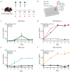
Qβ-VLPs based vaccine generation against candidate IAPP peptides. (a,b) Human IAPP amino acids (aa) sequence of the chosen peptides being coupled in a linear (a) and secondary (b) structure view: A N-terminal peptide without disulfide bond (N-term, in green); a N-terminal peptide with disulfide bond (s-s) between cystein 2 and 7 (N-term(s-s), in green and in red the disulphide bond)); a C-terminal peptide presents in the final 37-aa sequence (C-term, in light blue); and a C-terminal peptide (C-term-Pro, in orange) presents in the aa-sequence before final cleavage to the 37-aa long IAPP. (c) Firstly, Qβ-VLPs were chemically derivatized to heterobifunctional cross-linkers via Lys-aa (SMPH for the N-term, C-term and C-term-Pro peptides and s4FB for the N-term (s-s) peptide) and secondly coupled to the mentioned peptides via Cys-aa or an Aoa-group for the SMPH and the s4FB, respectively. The following vaccines were obtained: Qβ-Nterm (for coupling to N-term peptide without disulfide bond); Qβ-Nterm (s-s) (for coupling to the N-term (s-s) peptide with the disulfide bond); Qβ-Cterm, (for coupling to the C-term peptide); and Qβ-Cterm-Pro (for coupling to the C-term-Pro peptide). Coupling efficiency was analysed on a 15% SDS-PAGE under reducing condition. (d) To have detailed information of how many peptides per subunits were present, densitometry analysis was performed using Fiji ImageJ. The percent of the measured bands is displayed. Total area is represented as 100%. Qβ-VLPs, Virus-like particles derived from the bacteriophage Qβ; (s-s), disulphide bond; aa, amino acid; Lys, Lysin; Cys, Cystein, Aoa, aminooxyacetic group on the side chain of Lysin.
Figure 2
Figure 2
Immunogenicity control in C57BL/6 mice with the generated candidate vaccines. (a) Immunization scheme of 6 weeks old C57BL/6 mice. Mice were immunized at day 0 (d0), corresponding to the age of 6 weeks (w6). Boost was performed at day 14 (d14, w8) and day 28 (d28, w10). Blood was collected at day 0, day 14, day 28, day 42 and day 49 and serum was isolated. (b) 96-well ELISA plates were coated either with the corresponding peptide coupled on the vaccine used for immunization (in this case coupled to RNase), or with rat IAPP (rIAPP) or human IAPP (hIAPP). (cf) Total serum IgG of the mice receiving: the Qβ-Nterm vaccine (n = 4) (c) recognizing the RNase-Nterm peptide (in green); the Qβ-Nterm (s-s) vaccine (n = 4) recognizing the RNase-Nterm (s-s) peptide (in red); the Qβ-Cterm vaccine (n = 4) recognizing the RNase-Cterm peptide, and the Qβ-Cterm-Pro vaccine (n = 4) recognizing the RNase-Cterm-Pro peptide (in orange). Total IgG recognizing the hIAPP (square) and rIAPP (triangle) are shown in black. OD50 values are calculated as the corresponding dilution reaching to the half value of OD450. Data are the means ± SEM. d, day; w, week; rIAPP, rat IAPP; hIAPP, human IAPP; RNase-peptide, peptide covalently coupled to the corresponding peptide being either Nterm, Nterm (s-s), Cterm or Cterm-Pro.
Figure 3
Figure 3
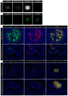
IgGs derived from C57BL/6 mice immunized with the Qβ-N-term (s-s) vaccine recognize only hIAPP aggregates on human and on hyperglycemic hIAPP transgenic mouse pancreatic tissue. (a) Nitrocellulose membrane coated either with rat IAPP (rIAPP), human IAPP monomers (hIAPP monomer), human IAPP fibrils (hIAPP fibrils) or a control amyloidogenic protein forming aggregates derived from amyloid beta (Aβ fibrils) followed incubation with purified polyclonal antibodies. After incubation with an anti-IgG conjugated to horseradish peroxidase, the membrane was developed with a chemiluminescent substrate. (b) Immediately after membrane blotting with the specified antigens, solutions were stained with a ThioflavinT (ThioT) solution and imaged to prove the state of the coated proteins. (c) Representative human pancreatic islet from a severe type 2 diabetes mellitus (T2DM) patients and a hyperglycemic transgenic mouse expressing hIAPP. Tissues were stained with ThioflavinS (ThioS) to confirm the presence of amyloidogenic aggregates (c, left panel, ThioS in green, DAPI in blue) and with an IgG derived from mice immunized with the Qβ-N-term(s-s) vaccine (c, middle panel, α-N-term (s-s) IgG in red, DAPI in blue). A co-localization staining (right panel, merge) was generated to authenticate the specificity of the IgGs. (d) Representative human pancreatic islet from a healthy pancreas and a wild-type mouse. ThioS staining (left panel) and polyclonal staining were checked on healthy human pancreatic tissue and wild-type normoglycemic mouse tissue. β-cells were visualized by staining with an anti-insulin (Ins, right panel) antibody. rIAPP, rat IAPP; hIAPP, human IAPP; Aβ, amyloid beta; α-N-term (s-s) IgG, polyclonal IgG derived from the Qβ-N-term (s-s) vaccinated C57BL/6 mice; ThioT, ThioflavinT; T2DM, type 2 diabetes mellitus; ThioS, ThioflavinS. Scale bars: 20 μm.
Figure 4
Figure 4
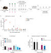
IAPP physiological function is maintained in the presence of anti-IAPP antibodies. (a) C57BL/6 male mice were immunized with the Qβ-Nterm (s-s) vaccine (n = 16) or with the uncoupled Qβ-VLPs (n = 14) at day 0 (week 6), day 14 (week 8) and day 28 (week 10), serum was collected at day 14 and day 34 (b) to check antibody titer. On days 36, 38, 41 and 43, mice were fasted during the light phase period for 12 h. Mice then received either IAPP or the control saline solution immediately prior to dark onset and food intake was measured 1, 2, 4 and 22 h after injection. (b) Serum antibodies were analyzed for recognition of the N-term (s-s) peptide coupled to RNase (RNase-N-term(s-s) in red), hIAPP (in peach) and rIAPP (in black) for the Qβ-Nterm (s-s) (left panel) and the Qβ-VLP (right panel)-immunized mice. (c) Cumulative food intake after 1-h refeeding and (d) baseline correction for the IAPP versus saline group in Qβ and Qβ-Nterm (s-s)-immunized mice. Additional time points can be seen in Supplementary Figure S2. Data are the means ± SEM. RNase-N-term (s-s), N-term (s-s) peptide coupled to RNase; rIAPP, rat IAPP; hIAPP, human IAPP. Statistical test in (c,d): 2-way ANOVA and Sidak’s multiple comparison. No significant difference was observed in (c,d) (p > 0.05).
Figure 5
Figure 5
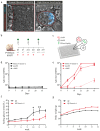
Immunization with the Qβ-Nterm(s-s) vaccine protects hIAPP transgenic mice from hyperglycemia and body weight loss. (a) Representative tomographic pictures merged with Thioflavin S (in blue) staining of pancreatic section of wild-type FVB and homozygous hyperglycemic male mice. Pancreatic islets are outlined in red. (b) Vaccination schedule of homozygous male FVB/Ins-hIAPP transgenic mice receiving either Qβ-Nterm (s-s) vaccine (n = 8) or uncoupled Qβ control (n = 7). (c) ELISA plates were coated either with the RNase-Nterm (s-s) peptide complex, with human IAPP (hIAPP), or with rat IAPP (rIAPP) to check for specific epitope recognition. (d) Total IgG titer of mice receiving the Qβ control. IgGs did not give positive signal for recognition of the RNase-Nterm (s-s) peptide complex (continuous black line), the hIAPP (dash black line) and the rIAPP (pointed black line). (e) Total IgG titer of mice receiving the Qβ-Nterm (s-s) vaccine. IgGs derived from the immunized mice recognized the RNase-Nterm (s-s) peptide complex (continuous red line), the hIAPP (dash red line) and the rIAPP (pointed red line). Qβ-immunized mice IgG did not give a positive signal for recognition of the RNase-Nterm (s-s) peptide complex (continuous black line), the hIAPP (dash black line) and the rIAPP (pointed black line). (f) Overnight fasting glycemia level and (g) body weight development in Qβ-Nterm (s-s) immunized (in red) and Qβ control group (in black). Statistical test; two-way ANOVA. Data are the means ± SEM. * p < 0.05, ** p < 0.01. ThioS, ThioflavS; (0/0), wildtype; (tg/tg), homozygous; RNase-N-term (s-s), N-term (s-s) peptide coupled to RNase; rIAPP, rat IAPP; hIAPP, human IAPP. Scale bars: 20 μm.
Figure 6
Figure 6
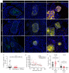
Immunization with the Qβ-Nterm(s-s) prevents fibrils depositions in pancreatic islets of hIAPP transgenic mice. (a,c,e) Paraffin-embedded pancreatic sections of 12-week-old Qβ (first row, Qβ, n = 32), Qβ-Nterm (s-s) (middle row, Qβ-Nterm (s-s), n = 28)-immunized homozygous male FVB/Ins-hIAPP transgenic mice and non-immunized wild-type FVB (last row, wildtype, n = 8) mice. (a) Fibril deposits were stained with ThioflavinS (ThioS, in green) and nuclei with DAPI (in blue). (b) Quantification of the relative amyloid/islet area in Qβ-Nterm (s-s) (in red), the Qβ control (in black)-immunized mice and the wild-type FVB control mice (in grey). (c) Insulin producing β-cells were stained with an anti-insulin antibody (in yellow) and nuclei with DAPI (in blue). (d) Correlation between the mean insulin intensity versus the pancreatic islet area of the Qβ control group (in black), the Qβ-Nterm (s-s)-immunized mice (in red) and the wild-type control (in grey). (e) Detection of IL-1β (in red), insulin (in yellow) in the pancreatic islets, and the nuclei with DAPI (in blue). (e) Magnification of the corresponding islet. (f) Quantifications of insulin-positive/total (left panel) and of the IL-1β-positive (right panel) pancreatic islet cells. Scale bars: 20 and 10 μm for the magnified pictures. Statistical test: Mann-Whitney test. Data are the means ± SEM. Ns: not significant, * p < 0.05, ** p < 0.01, *** p < 0.001, **** p < 0.0001. ThioS, ThioflavS; Ins, Insulin, IL-1β, Interleukin-1 β; wt, wild type.

Similar articles

Cited by

References

    1. Mathers C.D., Loncar D. Projections of global mortality and burden of disease from 2002 to 2030. PLoS Med. 2006;3:2011–2030. doi: 10.1371/journal.pmed.0030442. - DOI - PMC - PubMed
    1. Lascar N., Brown J., Pattison H., Barnett A.H., Bailey C.J., Bellaary S. Type 2 diabetes in adolescents and young adults. Lancet Diabetes Endocrinol. 2018;6:69–80. doi: 10.1016/S2213-8587(17)30186-9. - DOI - PubMed
    1. Kahn B.B. Type 2 Diabetes: When Insulin Minireview Secretion Fails to Compensate for Insulin Resistance docking proteins called insulin receptor substrates (IRSs). IRSs bind to various effector molecules including the regulatory subunit of phosphoinositol 3-kinase. Cell. 1998;92:593–596. doi: 10.1016/S0092-8674(00)81125-3. - DOI - PubMed
    1. Butler A.E., Janson J., Bonner-Weir S., Ritzel R., Rizza R.A., Butler P.C. β-Cell Deficit and Increased β-Cell Apoptosis in Humans With Type 2 Diabetes. Diabetes. 2003;52:102–110. doi: 10.2337/diabetes.52.1.102. - DOI - PubMed
    1. Westermark P. Quantitative Studies of Amyloid in the Islets of Langerhans. Ups. J. Med. Sci. 2016;77:91–94. doi: 10.1517/03009734000000014. - DOI - PubMed

Grants and funding