Review
. 2020 Mar 2;9(3):592.
doi: 10.3390/cells9030592.

Exosomes: From Potential Culprits to New Therapeutic Promise in the Setting of Cardiac Fibrosis

Affiliations
Review

Exosomes: From Potential Culprits to New Therapeutic Promise in the Setting of Cardiac Fibrosis

Roman Tikhomirov et al. Cells. .

Abstract

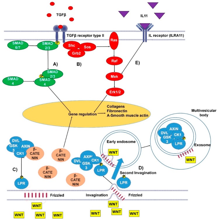
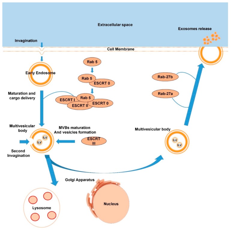
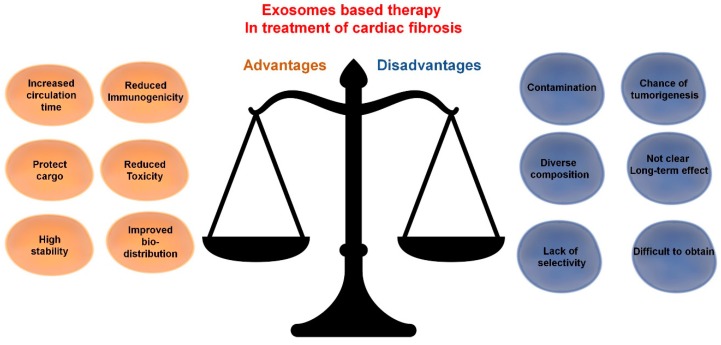
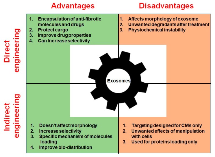
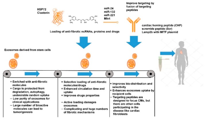
Fibrosis is a significant global health problem associated with many inflammatory and degenerative diseases affecting multiple organs, individually or simultaneously. Fibrosis develops when extracellular matrix (ECM) remodeling becomes excessive or uncontrolled and is associated with nearly all forms of heart disease. Cardiac fibroblasts and myofibroblasts are the main effectors of ECM deposition and scar formation. The heart is a complex multicellular organ, where the various resident cell types communicate between themselves and with cells of the blood and immune systems. Exosomes, which are small extracellular vesicles, (EVs), contribute to cell-to-cell communication and their pathophysiological relevance and therapeutic potential is emerging. Here, we will critically review the role of endogenous exosomes as possible fibrosis mediators and discuss the possibility of using stem cell-derived and/or engineered exosomes as anti-fibrotic agents.

Keywords: EVs engineering; cardiac fibrosis; exosomes; extracellular vesicle (EVs); heart failure; microRNAs; noncoding RNAs; stem cells.


Conflict of interest statement

The authors declare that there is no conflict of interest regarding the publication of this article.

Figures

Figure 1
Figure 1
The canonical and non-canonical pro-fibrotic pathways of transforming growth factor β (TGFβ) and Wnt and the pro-fibrotic interleukin 11 (IL-11) pathway. (A) Canonical TGFβ pathway: TGFβ binds to the type I/II TGFβ receptor. The Smad2/3 complex is then phosphorylated, at this point the pathway can be inhibited by Smad6/7. In the cytoplasm Smad2/3 binds Smad4 and the whole complex transfers to the nucleus, where it binds to GAGAC motifs, promoting gene expression. (B) Non-canonical TGFβ pathway: TGFβ binds the TGFβ receptor type I/II, resulting in tyrosine residues and Shc (Src homology 2 domain containing) transforming protein phosphorylation. This promotes the binding of Grb2 (Growth factor receptor-bound protein 2) and Sos (son of sevenless). This complex can activate Ras, launching the MAPK cascade and further gene regulation. (C) Wnt canonical pathway: Wnt (wingless-related integration site) binds transmembrane protein frizzled (Fz). Fz bound WNT can then bind LPR5/6 protein (low-density-lipoprotein-related protein) which undergoes phosphorylation of its tail by GSK3 and CK1 proteins. Following this, low-density-lipoprotein receptor-related protein (LRP) interacts with Disheveld (DVL), Axin, and GSK3 via Pro-Pro-Pro-(Ser/Tyr)-Pro repeats. This complex is responsible for β-catenin attenuation. (D) Wnt non-canonical pathway: Wnt binds transmembrane protein Fz at the site of a membrane invagination. The complex then becomes part of the membrane of an early endosome. LPR5/6 is phosphorylated at its tail by GSK3 and CK1 proteins. Following this, LRP interacts with DVL, Axin, and GSK3 via Pro-Pro-Pro-(Ser/Tyr)-Pro repeats. The membrane of the early endosome can form a second invagination, eventually leading to the whole complex being locked down inside a multivesicular body. (E) The IL-11 signaling pathway activates the MAPK cascade, promoting gene regulation [15,16,24].
Figure 2
Figure 2
Scheme of exosome biogenesis (the process is described in detail in Section 1.2.1.).
Figure 3
Figure 3
Exosome-based therapy in treatment of cardiac fibrosis: advantages and disadvantages.
Figure 4
Figure 4
Advantages and disadvantages of direct and indirect exosome engineering.
Figure 5
Figure 5
Current benefits and limitations of exosome-based therapies in the treatment of cardiac fibrosis.

References

    1. Murtha L.A., Schuliga M.J., Mabotuwana N.S., Hardy S.A., Waters D.W., Burgess J.K., Knight D.A., Boyle A.J. The Processes and Mechanisms of Cardiac and Pulmonary Fibrosis. Front. Physiol. 2017;8:777. doi: 10.3389/fphys.2017.00777. - DOI - PMC - PubMed
    1. Ma C.X., Zhao X.K., Li Y.D. New therapeutic insights into radiation-induced myocardial fibrosis. Ther. Adv. Chronic Dis. 2019;10:2040622319868383. doi: 10.1177/2040622319868383. - DOI - PMC - PubMed
    1. Gulati A., Japp A.G., Raza S., Halliday B.P., Jones D.A., Newsome S., Ismail N.A., Morarji K., Khwaja J., Spath N., et al. Absence of Myocardial Fibrosis Predicts Favorable Long-Term Survival in NewOnset Heart Failure. Circulation. 2018;11:e007722. - PubMed
    1. Tian J., An X., Niu L. Myocardial fibrosis in congenital and pediatric heart disease. Exp. Ther. Med. 2017;13:1660–1664. doi: 10.3892/etm.2017.4224. - DOI - PMC - PubMed
    1. Ytrehus K., Hulot J.S., Perrino C., Schiattarella G.G., Madonna R. Perivascular fibrosis and the microvasculature of the heart. Still hidden secrets of pathophysiology? Vasc. Pharm. 2018;107:78–83. doi: 10.1016/j.vph.2018.04.007. - DOI - PubMed

Publication types