

Ginseng Gintonin Attenuates Lead-Induced Rat Cerebellar Impairments during Gestation and Lactation
- PMID: 32131481
- PMCID: PMC7175158
- DOI: 10.3390/biom10030385
Ginseng Gintonin Attenuates Lead-Induced Rat Cerebellar Impairments during Gestation and Lactation
Abstract
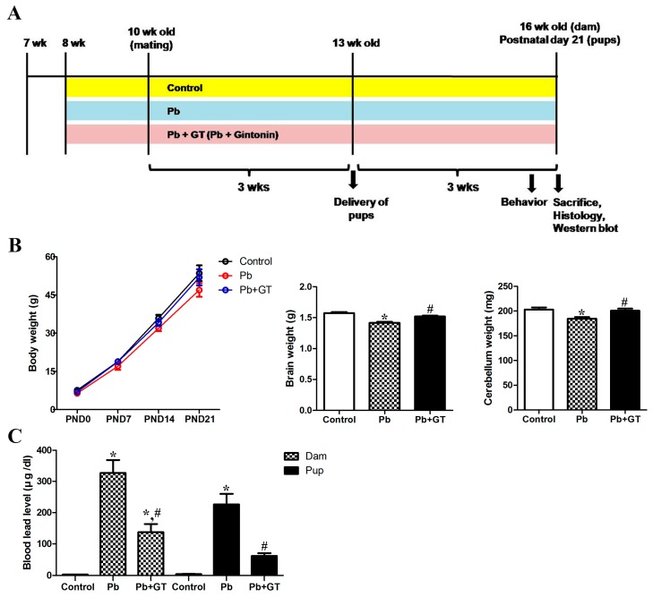
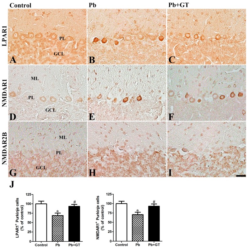
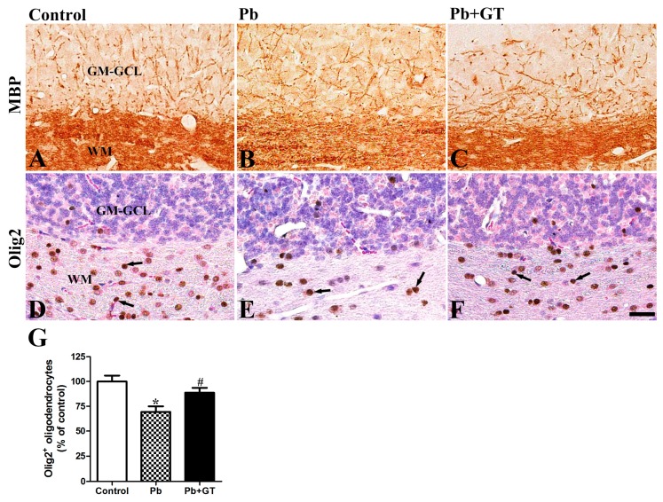
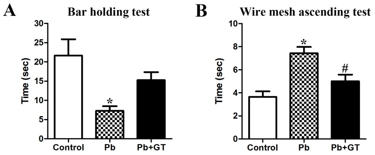
Gintonin, a novel ginseng-derived lysophosphatidic acid receptor ligand, improves brain functions and protects neurons from oxidative stress. However, little is known about the effects of gintonin against Pb-induced brain maldevelopment. We investigated the protective effects of gintonin on the developing cerebellum after prenatal and postnatal Pb exposure. Pregnant female rats were randomly divided into three groups: control, Pb (0.3% Pb acetate in drinking water), and Pb plus gintonin (100 mg/kg, p.o.). Blood Pb was increased in dams and pups; gintonin treatment significantly decreased blood Pb. On postnatal day 21, the number of degenerating Purkinje cells was remarkably increased while the number of calbindin-, GAD67-, NMDAR1-, LPAR1-immunoreactive intact Purkinje cells, and GABA transporter 1-immunoreactive pinceau structures were significantly reduced in Pb-exposed offspring. Following Pb exposure, gintonin ameliorated cerebellar degenerative effects, restored increased pro-apoptotic Bax, and decreased anti-apoptotic Bcl2. Gintonin treatment attenuated Pb-induced accumulation of oxidative stress (Nrf2 and Mn-SOD) and inflammation (IL-1β and TNFα,), restoring the decreased cerebellar BDNF and Sirt1. Gintonin ameliorated Pb-induced impairment of myelin basic protein-immunoreactive myelinated fibers of Purkinje cells. Gintonin attenuated Pb-induced locomotor dysfunctions. The present study revealed the ameliorating effects of gintonin against Pb, suggesting the potential use of gintonin as a preventive agent in Pb poisoning during pregnancy and lactation.
Keywords: cerebellar lead (Pb) poisoning; ginseng; gintonin; neuroprotection; pregnancy and lactation.
Conflict of interest statement
The authors declare no conflict of interest.
Figures


Similar articles
-
Effects of ascorbic acid treatment on developmental alterations in calcium-binding proteins and gamma-aminobutyric acid transporter 1 in the cerebellum of lead-exposed rats during pregnancy and lactation.J Toxicol Sci. 2019;44(11):799-809. doi: 10.2131/jts.44.799. J Toxicol Sci. 2019. PMID: 31708536
-
Ascorbic Acid Supplementation Prevents the Detrimental Effects of Prenatal and Postnatal Lead Exposure on the Purkinje Cell and Related Proteins in the Cerebellum of Developing Rats.Biol Trace Elem Res. 2019 Aug;190(2):446-456. doi: 10.1007/s12011-018-1572-y. Epub 2018 Nov 28. Biol Trace Elem Res. 2019. PMID: 30488169
-
Protective effect of garlic extract against maternal and foetal cerebellar damage induced by lead administration during pregnancy in rats.Folia Morphol (Warsz). 2018;77(1):1-15. doi: 10.5603/FM.a2017.0063. Epub 2017 Jul 13. Folia Morphol (Warsz). 2018. PMID: 28703846
-
Gintonin: a novel ginseng-derived ligand that targets G protein- coupled lysophosphatidic acid receptors.Curr Drug Targets. 2012 Dec;13(13):1659-64. doi: 10.2174/138945012803529947. Curr Drug Targets. 2012. PMID: 23017203 Review.
-
Ginseng gintonin, aging societies, and geriatric brain diseases.Integr Med Res. 2021 Mar;10(1):100450. doi: 10.1016/j.imr.2020.100450. Epub 2020 Jun 13. Integr Med Res. 2021. PMID: 32817818 Free PMC article. Review.
Cited by
-
Modulation of Kynurenic Acid Production by N-acetylcysteine Prevents Cognitive Impairment in Adulthood Induced by Lead Exposure during Lactation in Mice.Antioxidants (Basel). 2023 Nov 23;12(12):2035. doi: 10.3390/antiox12122035. Antioxidants (Basel). 2023. PMID: 38136155 Free PMC article.
-
Combining in vitro assays and mathematical modelling to study developmental neurotoxicity induced by chemical mixtures.Reprod Toxicol. 2021 Oct;105:101-119. doi: 10.1016/j.reprotox.2021.08.007. Epub 2021 Aug 26. Reprod Toxicol. 2021. PMID: 34455033 Free PMC article.
-
Effects of Gintonin-enriched fraction on the gene expression of six lysophosphatidic receptor subtypes.J Ginseng Res. 2021 Sep;45(5):583-590. doi: 10.1016/j.jgr.2021.02.006. Epub 2021 Feb 22. J Ginseng Res. 2021. PMID: 34803428 Free PMC article.
-
Investigating the protective properties of Panax ginseng and its constituents against biotoxins and metal toxicity: a mechanistic review.Naunyn Schmiedebergs Arch Pharmacol. 2025 Feb;398(2):1215-1242. doi: 10.1007/s00210-024-03410-2. Epub 2024 Sep 17. Naunyn Schmiedebergs Arch Pharmacol. 2025. PMID: 39287674 Review.
-
Topical Collection "Pharmacology of Medicinal Plants".Biomolecules. 2021 Jan 14;11(1):101. doi: 10.3390/biom11010101. Biomolecules. 2021. PMID: 33466709 Free PMC article.
References
-
- Kim H.J., Kim D.J., Shin E.J., Lee B.H., Choi S.H., Hwang S.H., Rhim H., Cho I.H., Kim H.C., Nah S.Y. Effects of gintonin-enriched fraction on hippocampal cell proliferation in wild-type mice and an APPswe/PSEN-1 double Tg mouse model of Alzheimer’s disease. Neurochem. Int. 2016;101:56–65. doi: 10.1016/j.neuint.2016.10.006. - DOI - PubMed
-
- Jang M., Choi J.H., Chang Y., Lee S.J., Nah S.Y., Cho I.H. Gintonin, a ginseng-derived ingredient, as a novel therapeutic strategy for Huntington’s disease: Activation of the Nrf2 pathway through lysophosphatidic acid receptors. Brain Behav. Immu. 2019;80:146–162. doi: 10.1016/j.bbi.2019.03.001. - DOI - PubMed
-
- Lee M.J., Chang B.J., Oh S., Nah S.Y., Cho I.H. Korean red ginseng mitigates spinal demyelination in a model of acute multiple sclerosis by downregulating p38 mitogen-activated protein kinase and nuclear factor-κB signaling pathways. J. Ginseng Res. 2018;42:436–446. doi: 10.1016/j.jgr.2017.04.013. - DOI - PMC - PubMed
Publication types
MeSH terms
Substances
LinkOut - more resources
Full Text Sources
Medical
Research Materials
