. 2021 May;31(4):272-287.
doi: 10.1080/15376516.2020.1736702. Epub 2020 Mar 30.

Halogen gas exposure: toxic effects on the parturient

Affiliations

Halogen gas exposure: toxic effects on the parturient

Dylan R Addis et al. Toxicol Mech Methods. 2021 May.

Abstract

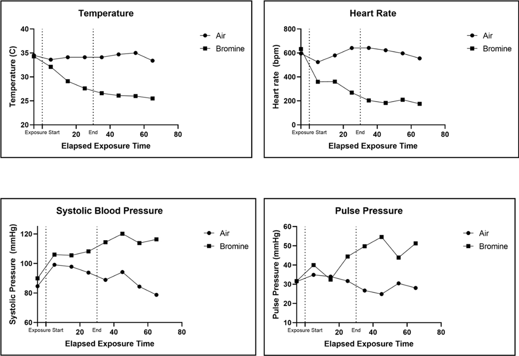
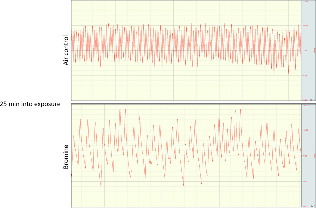
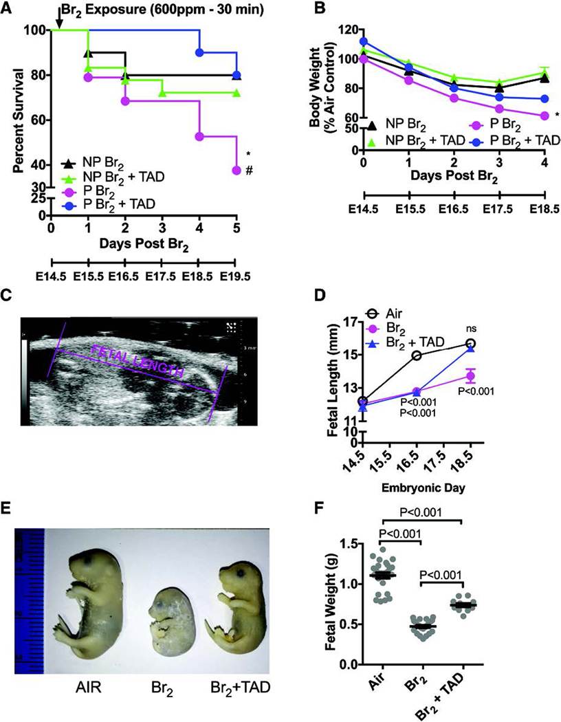
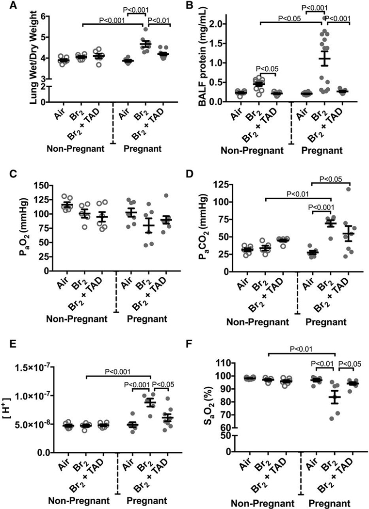
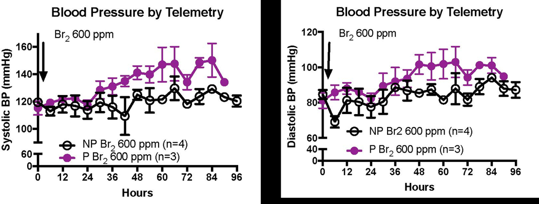
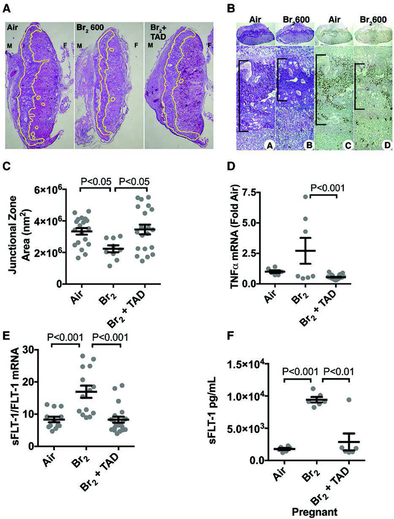
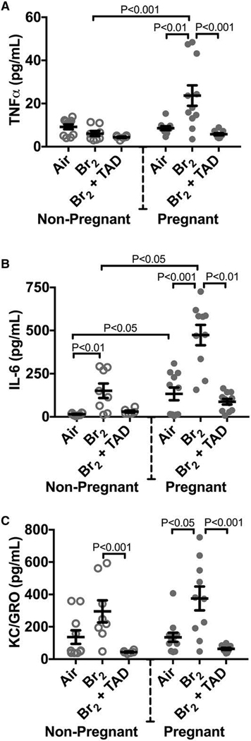
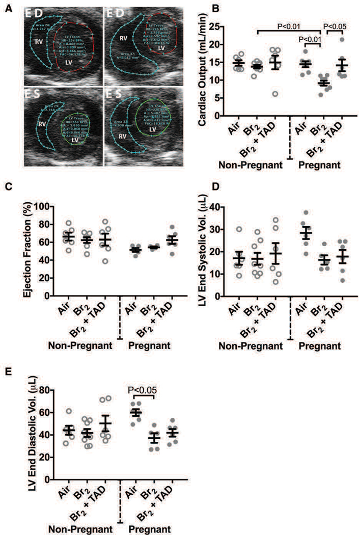
The elemental halogens include chlorine, bromine, and phosgene. Halogen gas can be directly weaponized and employed in warfare or terrorism. Industrial stockpiles or halogen transport can provide targets for terrorist attack as well as an origin for accidental release creating a risk for potential mass-casualty incidents. Pregnant and post-partum women represent a substantial and vulnerable subset of the population who may be at particular risk during an attack or accidental exposure. We review the effects of halogen exposure on the parturient with a focus on bromine toxicity. Bromine is the most extensively studied agent in the context of pregnancy and to-date murine models form the basis for the majority of current knowledge. Pregnancy potentiates the acute lung injury after halogen exposure. In addition, halogen exposure precipitates a preeclamptic-like syndrome in mice. This phenotype is characterized by systemic and pulmonary hypertension, endothelial dysfunction, decreased cardiac output, placental injury and fetal growth restriction. This constellation contributes to increased maternal and fetal mortality observed after bromine exposure. Angiogenic imbalance is noted with overexpression of the soluble fms-like tyrosine kinase-1 (sFlt-1) form of the vascular endothelial growth factor receptor 1 reminiscent of human preeclampsia. Additional research is needed to further explore the effect of halogen gas exposure in pregnancy and to develop therapeutic interventions to mitigate risk to this unique population.

Keywords: Halogen; bromine; cardiopulmonary injury; chlorine; pregnancy.


Figures

Figure 1.
Figure 1.
Hemodynamic data obtained from telemetry recordings in two mice exposed to either air or Br2 (600 ppm for 30 min). Dashed lines demarcate the beginning and end of Br2 exposure. The telemetry pressure transducer was located in the aortic arch. Br2: Bromine
Figure 2.
Figure 2.
Representative telemetry aortic arch pressure tracings obtained during exposure to bromine (600 ppm) or air at 25 minutes into a 30 minute exposure. Bradycardia, an increased systolic blood pressure, and a widened pulse pressure are evident.
Figure 3.
Figure 3.. Pregnant mice exhibit increased body weight loss and mortality and fetal growth restriction.
Non-pregnant (NP) and pregnant (P) (E14.5) mice were exposed to air or to Br2 at 600 ppm for 30 min and returned to room air; they received tadalafil (TAD; 2 mg/kg BW in 0.1 ml of sterile saline) or vehicle via oral gavage at 1 h post-exposure and every 24 h thereafter. Body weights and survival times were recorded daily. A) Kaplan-Meyer curves of pregnant and non-pregnant mice with tadalafil or vehicle, post Br2 exposure. Non-pregnant mice exposed to Br2 and returned to room air lived longer than similarly exposed pregnant mice (* = P<0.05). Tadalafil improved survival times of pregnant mice post Br2 (# = P<0.05) but not of non-pregnant mice; n=10–19; Log-Rank Test. B) Body weights normalized to weights of air-exposed mice. Pregnant mice exposed to Br2 and returned to room air exhibit more severe weight loss compared to similarly exposed non-pregnant mice (* = P<0.05). Tadalafil administration mitigated weight loss in pregnant mice at four days post-exposure, but has no effect in non-pregnant mice; n=6–8; ANOVA. C) Representative ultrasound of a fetus showing how fetal length was measured. D) Summary data of fetal length measurements at E14.5, E16.5, & E18.5. Exposure to Br2 resulted in decreased fetal lengths which were restored to their air control values at E18.5 in the tadalafil group; n= 6–10 pups (2 pups per litter) for each condition; ANOVA; p values as compared to the corresponding air controls for the indicated gestational age. E) Representative photograph of paraformaldehyde-fixed fetuses at E18.5 for the indicated conditions. Fetuses of Br2-exposed pregnant mice exhibit severe fetal growth restriction, and tadalafil improves fetal growth. F) Fetal weights were recorded after extraction of fetuses at E18.5. Fetal weights of fetuses from Br2-exposed pregnant mice weighed considerably less and were partially rescued by tadalafil (TAD); n=pups (11–25) (2 pups per litter); ANOVA. All data are means±S.E.M.
Figure 4.
Figure 4.. Pregnant mice exhibit pulmonary injury 96 h post-exposure.
Non-pregnant and pregnant (E14.5) mice were exposed to air or 600ppm Br2 for 30 minutes, returned to room air, then administered tadalafil (TAD) or vehicle. A) Lung wet/dry weight ratios at E18.5 are increased in pregnant mice exposed to Br2 and are reduced by tadalafil. B) Protein concentrations in the bronchoalveolar lavage fluid (BALF) of pregnant mice were increased at E18.5 and returned to the air control values following administration of tadalafil (TAD). C–F) PaO2 was unchanged, PaCO2 increased, [H+] increased and SaO2 was decreased in pregnant mice exposed to Br2 at E18.5. Administration of Tadalafil returned [H+] and SaO2 to their air control values but did not improve PaCO2; All data n=6–14 mice; ANOVA; means±S.E.M.
Figure 5.
Figure 5.
Levels of brominated fatty acids (18C = stearic acid) in the plasma of non-pregnant or pregnant mice at 1 hour post exposure to bromine (600 ppm for 30 min). Individual values for each mouse along with means ± SEM. There was no difference among the means of pregnant and non-pregnant mice for 18C brominated fatty acid. ANOVA, Tukey’s multiple comparisons test. n=6–8. NP: non-pregnant, P: pregnant, C: carbon.
Figure 6.
Figure 6.. Exposure to Br2 increases the systemic blood pressure of pregnant mice.
Non-pregnant and pregnant (E14.5) mice were exposed to air or 600ppm Br2 for 30 minutes then returned to room air. At E18.5, a pressure-transducer catheter was inserted into the aortic arch via the carotid in anesthetized mice. Increases in both systolic and diastolic pressure were noted in pregnant mice.
Figure 7.
Figure 7.. Exposure of pregnant mice to Br2 damages their placentas.
Pregnant (E14.5) mice were exposed to air or 600ppm Br2 for 30 minutes, returned to room air and received tadalafil (TAD) or vehicle. A–B) Representative H&E stained (A) placenta sections at E18.5 with the junctional zone demarcated with yellow highlighting (A) as well as (B) PAS staining (left) and CDX2 staining (right) of Br2-exposed pregnant mice revealed a reduced junctional zone (B: black bars) at E18.5; tadalafil administration restored junctional zones to normal size. C) Junctional zone areas at E18.5 for the indicated groups; n=9–20; ANOVA. D–E) TNFα mRNA (n=6–23) was reduced in Br2-exposed pregnant mice treated with tadalafil. sFLT-1/FLT-1 mRNA (=12–18) was increased in placentas of Br2-exposed pregnant mice at E18.5 (ANOVA), and was reduced to air control values by tadalafil. F) sFLT-1 in plasma at E18.5 increased in pregnant mice exposed to Br2 and was reduced with tadalafil; ANOVA. Only one placenta per pregnant mother was used. All data are means±S.E.M.
Figure 8.
Figure 8.
Br2-exposed pregnant mice exhibit progressively increased circulating sFLT-1 (soluble fms-like tyrosine kinase 1) with a concomitant increase of lung wet/dry weight. Non-pregnant and pregnant (E14.5) mice were exposed to air or to Br2 at 600 ppm for 30 minutes and returned to room air. sFLT-1 levels in the plasma of (A) pregnant Br2-exposed mice increased 48-hour post-exposure vs air controls and continued to increase until euthanasia at 96 hours. This increase was linear (R2=0.8151, P<0.0001). Similarly exposed (B) non-pregnant mice exhibited no increase at any time point; n=6 to 8; ANOVA. Lung wet:dry weight ratios of (C) pregnant Br2-exposed mice increased 72-hour post-exposure compared with air controls and continued to increase until euthanasia at 96 hours. Similarly exposed (D) non-pregnant mice exhibited no increase at any time point; n=6 to 8; ANOVA. All data are individual values and means±SEM. sFLT-1 indicates soluble fms-like tyrosine kinase 1.
Figure 9.
Figure 9.. Br2-exposed pregnant mice exhibit diminished cardiac function at E18.5.
Non-pregnant and pregnant (E14.5) mice were exposed to air or 600ppm Br2 for 30 minutes, returned to room air, then administered tadalafil (TAD) or vehicle. A) Representative echosonography LV and RV traces at E18.5 of pregnant mice exposed to air or Br2 with demarcation of ventricular sizes. HR = Heart rate, A = Area, A;s = Area systole, A;d = Area diastole, FAC = Fractional area change. B) Cardiac output (LV) determined by echosonography was decreased in Br2-exposed pregnant mice, which increased following administration of tadalafil; ANOVA: n=6–8. C) Ejection fraction (LV) was unchanged in all groups; ANOVA. D) LV end-systolic volume was similar in all groups. E) However, LV end-diastolic volume was diminished in Br2-exposed pregnant mice; n=6–8; ANOVA. All data are means±S.E.M.
Figure 10.
Figure 10.. Pregnant mice exposed to Br2 demonstrated systemic inflammation at E18.5.
Non-pregnant and pregnant (E14.5) mice were exposed to air or 600ppm Br2 for 30 minutes, returned to room air, then administered tadalafil (TAD) or vehicle. A–C) Plasma TNFα, IL-6, and KC/GRO increased in pregnant mice exposed to Br2 but only IL-6 increased in non-pregnant mice post-Br2. All three cytokines in pregnant mice exposed to Br2 returned to air control values following tadalafil administration; n=6–12 per group; ANOVA. All data are means±S.E.M.

References

    1. Addis DR, Lambert JA, Ren C, Doran S, Aggarwal S, Jilling T, Matalon S. 2020. Vascular Endothelial Growth Factor-121 Administration Mitigates Halogen Inhalation-Induced Pulmonary Injury and Fetal Growth Restriction in Pregnant Mice. Journal of the American Heart Association. 9(3):e013238. - PMC - PubMed
    1. Aggarwal S, Ahmad I, Lam A, Carlisle MA, Li C, Wells JM, Raju SV, Athar M, Rowe SM, Dransfield MT et al. 2018. Heme scavenging reduces pulmonary endoplasmic reticulum stress, fibrosis, and emphysema. JCI Insight. 3(21). - PMC - PubMed
    1. Aggarwal S, Lam A, Bolisetty S, Carlisle MA, Traylor A, Agarwal A, Matalon S. 2016. Heme Attenuation Ameliorates Irritant Gas Inhalation-Induced Acute Lung Injury. Antioxid Redox Signal. 24(2):99–112. - PMC - PubMed
    1. Ahmad S, Ahmad A, Hendry-Hofer TB, Loader JE, Claycomb WC, Mozziconacci O, Schoneich C, Reisdorph N, Powell RL, Chandler JD et al. 2015. Sarcoendoplasmic reticulum Ca(2+) ATPase. A critical target in chlorine inhalation-induced cardiotoxicity. Am J Respir Cell Mol Biol. 52(4):492–502. - PMC - PubMed
    1. Ahmad S, Masjoan Juncos JX, Ahmad A, Zaky A, Wei CC, Bradley WE, Zafar I, Powell P, Mariappan N, Vetal N et al. 2019. Bromine inhalation mimics ischemia-reperfusion cardiomyocyte injury and calpain activation in rats. Am J Physiol Heart Circ Physiol. 316(1):H212–h223. - PMC - PubMed

Substances