. 2020 Mar 4;15(1):87.
doi: 10.1186/s13018-020-01595-9.

The JNK pathway represents a novel target in the treatment of rheumatoid arthritis through the suppression of MMP-3

Affiliations

The JNK pathway represents a novel target in the treatment of rheumatoid arthritis through the suppression of MMP-3

Tomotake Kanai et al. J Orthop Surg Res. .

Abstract

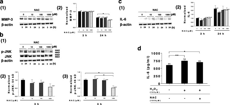
Background and aim: The pathophysiology of rheumatoid arthritis (RA) is characterized by excess production of pro-inflammatory cytokines, including tumor necrosis factor-α (TNF-α), interleukin-1β (IL-1β), and interleukin-6 (IL-6) by neutrophils and macrophages in synovium. Additionally, these cytokines promote the production of reactive oxygen species (ROS), and increased production of matrix metalloproteinases (MMPs), including MMP-3, in synoviocytes that result in joint destruction. There is limited information on how proteolytic enzymes such as MMP-3 can be regulated. We evaluated the effect of the antioxidant N-acetylcysteine (NAC) on RA and identified the relationship between the c-Jun N terminal kinase (JNK) pathway and MMP-3. We hypothesized that elucidating this relationship would lead to novel therapeutic approaches to RA treatment and management.

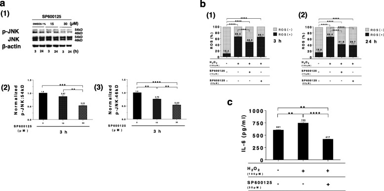
Methods: We investigated the effect of administering a low dose (1000 μM or less) of an antioxidant (NAC) to human rheumatoid fibroblast-like synoviocytes (MH7A cells). We also investigated the response of antioxidant genes such as nuclear factor erythroid -derived 2-related factor 2 (Nrf2) and Sequestosome 1 (p62). The influence of MMP-3 expression on the JNK pathway leading to joint destruction and the mechanisms underlying this relationship were investigated through primary dispersion culture cells collected from the synovial membranes of RA patients, consisting of rheumatoid arthritis-fibroblast-like synoviocytes (RA-FLS).

Results: Low-dose NAC (1000 μM) increased the expression of Nrf2 and phospho-p62 in MH7A cells, activating antioxidant genes, suppressing the expression of MMP-3, and inhibiting the phosphorylation of JNK. ROS, MMP-3 expression, and IL-6 was suppressed by administering 30 μM of SP600125 (a JNK inhibitor) in MH7A cells. Furthermore, the administration of SP600125 (30 μM) to RA-FLS suppressed MMP-3.

Conclusions: We demonstrated the existence of an MMP-3 suppression mechanism that utilizes the JNK pathway in RA-FLS. We consider that the JNK pathway could be a target for future RA therapies.

Keywords: JNK pathway; Low-dose NAC; MH7A; MMP-3; RA-FLS.


Conflict of interest statement

The authors declare that they have no competing interest.

Figures

Fig. 1
Fig. 1
Anti-oxidative effects of NAC. a(1) Cell viability graphs following NAC treatment at concentrations of 10, 100, 500, 1000 μM, and 10 mM for 24 h. All data are expressed as the mean ± SEM (N = 4). The dash-dotted line is a 10 mM NAC concentration producing 30% cell viability. a(2) This graph is an enlargement of a(1) below 1000 μM. The concentration of NAC showing less than 10% cytotoxicity was 1000 μM. a(3) The cell viability assay of H2O2. H2O2 more than 100 μM was cytotoxic, killing 60% of cells in the sample. b(1), c(1) An illustrative Western blotting picture of Nrf2 and phospho-p62 (p-p62) following NAC treatment (1000 μM) for 3 or 24 h. b(2), c(2) Normalized values of Nrf2 or p-p62 were graphed (N = 3). Normalization was performed with Nrf2 or p-p62 band intensity of untreated cell (3-h incubation). d Immunocytochemistry of MH7A cells stained to identify Nrf2. The color analyzed by confocal laser scanning microscopy is expressed as Nrf2 (green)/phalloidin (red)/DAPI (blue). d(1) Untreated cells were cultured for 24 h. d(2) Cells cultured for 24 h treated with low-dose NAC (1000 μM). Scale bar; 20 μm. e ROS formation in MH7A cells following H2O2 treatment (100 μM) for 1 h and low-dose NAC treatment (1000 μM) for 3 (e(1)) and 24 h (e(2)). All values are expressed as the mean ± SEM (N = 3). The significance levels are shown as *P value < .05, **P value < .01, ***P value < .001, ****P value < .0001
Fig. 2
Fig. 2
Low-dose NAC suppressed MMP-3 expression and JNK phosphorylation. a(1) An illustrative Western blot picture of MMP-3 following low-dose NAC treatment (1000 μM) after 3 and 24 h. a(2) Normalized intensities were graphed (N = 3). Normalization was performed with MMP-3 band intensity of untreated MH7A cell (3-h incubation). b(1) An illustrative Western blot picture of phosphorylated JNK (54 and 46 kD) in MH7A cells following NAC (10, 100, and 1000 μM) for 3 and 24 h. b(2, 3) Normalized intensities were graphed (N = 3). Normalization was performed with phosphorylated JNK (p-JNK; 54 and 46 kD) of untreated MH7A cell (3-h incubation). c(1) An illustrative Western blot picture of IL-6 in MH7A cells following low-dose NAC treatment (1000 μM) after 3 or 24 h. c(2) Normalized intensities were graphed (N = 3). Normalization was performed with IL-6 bad intensity of untreated MH7A cell (3-h incubation). d This graph showed the mean concentrations of IL-6 in supernatant of MH7A cells determined by chemiluminescent enzyme immunoassay (CLEIA) following 24 h after H2O2 treatment (100 μM) and low-dose NAC treatment (1000 μM). All values are expressed as the mean ± SEM (N = 3). The significance levels are shown as *P value < .05, **P value < .01
Fig. 3
Fig. 3
JNK inhibitor (SP600125) has anti-oxidative and anti-inflammatory effects. a(1) An illustrative Western blot picture of phosphorylated JNK (54 and 46 kD) in MH7A cells following SP600125 treatment (15 and 30 μM) for 3 h. a(2, 3) Normalized intensities were graphed (N = 3). Normalization was performed with phosphorylated JNK (54 and 46 kD) of untreated MH7A cell (3-h incubation). b(1, 2) ROS formation assay in MH7A cells by H2O2 treatment (100 μM) for 1 h following SP600125 treatment (15 and 30 μM) for 3 and 24 h was evaluated by Muse Cell Analyzer. c This graph showed the mean concentrations of IL-6 in supernatant of MH7A cells determined by chemiluminescent enzyme immunoassay (CLEIA) following 24 h after the H2O2 treatment (100 μM) and SP600125 (30 μM). All values are expressed as the mean ± SEM (N = 3). The significance levels are shown as *P value < .05, **P value < .01, ***P value < .001, ****P value < .0001
Fig. 4
Fig. 4
JNK inhibitor suppresses MMP-3 in both MH7A cells and RA-FLS. a(1) An illustrative Western blot picture of MMP-3 following SP600125 treatment (15 and 30 μM) after 3 and 24 h in MH7A cells. a(2) Normalized intensities were graphed (N = 3). Normalization was performed with MMP-3 band intensity of untreated MH7A cell (3-h incubation). All values are expressed as the mean ± SEM (N = 3). The significance levels are shown as *P value < .05. b(1) An illustrative Western blot picture of MMP-3 following SP600125 treatment (15 and 30 μM) after 3 and 24 h in RA-FLS. b(2) Normalized intensities were graphed (N = 3). Normalization was performed with MMP-3 bad intensity of untreated RA-FLS cell (3-h incubation). All values are expressed as the mean ± SEM (N = 3). The significance levels are shown as *P value < .05
Fig. 5
Fig. 5
Schema of MMP-3 suppression via the JNK pathway by low-dose NAC and JNK inhibitor

Similar articles

Cited by

References

    1. Filippin LI, Vercelino R, Marroni NP, Xavier RM. Redox signaling and the inflammatory response in rheumatoid arthritis. Clin Exp Immunol. 2008;152:415–422. doi: 10.1111/j.1365-2249.2008.03634.x. - DOI - PMC - PubMed
    1. Kobayashi M, Squires GR, Mousa A, Tanzer M, Zukour D, Antoniou J, Feige U, Poole R. Role of interleukin-1 and tumor necrosis factor α in matrix degradation of human osteoarthritic cartilage. Arthritis Rhum. 2005;52:128–135. doi: 10.1002/art.20776. - DOI - PubMed
    1. Quinonez-Flores CM, Gonzalez Chavez SA, Najera DDR, Pacheco-Tena C. Oxidative stress relevance in the pathogenesis of the rheumatoid arthritis: a systematic review. Bio Med Res Int. 2016;6097417:1–14. - PMC - PubMed
    1. Zafarullah M, Li WQ, Sylvester J, Ahmad M. Molecular mechanisms of N-acetylcysteine actions. CMLS. 2003;60:6–20. doi: 10.1007/s000180300001. - DOI - PMC - PubMed
    1. Li J, Li J, Yue Y, Hu Y, Cheng W, Liu R, Pan X, Zhang P. Genistein suppresses tumor necrosis factor α-induced inflammation via modulating reactive oxygen species/Akt/nuclear factor κB and adenosine monophosphate-activated protein kinase signal pathways in human synoviocyte MH7A cells. Drug Des Devel Ther. 2014;8:315–323. doi: 10.2147/DDDT.S52354. - DOI - PMC - PubMed

MeSH terms