

Comparison of amyloid PET measured in Centiloid units with neuropathological findings in Alzheimer's disease
- PMID: 32131891
- PMCID: PMC7057642
- DOI: 10.1186/s13195-020-00587-5
Comparison of amyloid PET measured in Centiloid units with neuropathological findings in Alzheimer's disease
Abstract
Background: The Centiloid scale was developed to standardise the results of beta-amyloid (Aβ) PET. We aimed to determine the Centiloid unit (CL) thresholds for CERAD sparse and moderate-density neuritic plaques, Alzheimer's disease neuropathologic change (ADNC) score of intermediate or high probability of Alzheimer's Disease (AD), final clinicopathological diagnosis of AD, and expert visual read of a positive Aβ PET scan.
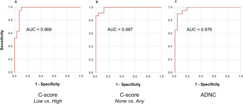
Methods: Aβ PET results in CL for 49 subjects were compared with post-mortem findings, visual read, and final clinicopathological diagnosis. The Youden Index was used to determine the optimal CL thresholds from receiver operator characteristic (ROC) curves.
Results: A threshold of 20.1 CL (21.3 CL when corrected for time to death, AUC 0.97) yielded highest accuracy in detecting moderate or frequent plaque density while < 10 CL was optimal for excluding neuritic plaque. The threshold for ADNC intermediate or high likelihood AD was 49.4 CL (AUC 0.98). Those cases with a final clinicopathological diagnosis of AD yielded a median CL result of 87.7 (IQR ± 42.2) with 94% > 45 CL. Positive visual read agreed highly with results > 26 CL.
Conclusions: Centiloid values < 10 accurately reflected the absence of any neuritic plaque and > 20 CL indicated the presence of at least moderate plaque density, but approximately 50 CL or more best confirmed both neuropathological and clinicopathological diagnosis of Alzheimer's disease.
Keywords: Alzheimer’s disease; Amyloid imaging; Centiloids; Neuropathology; Positron emission tomography.
Conflict of interest statement
Dr. Amadoru is site principal investigator for the AbbVie ABBV-8E12 AWARE study at Austin Health. He receives a government hospital award wage for his Austin Health clinical and research appointments.
Dr. Doré reports no relevant disclosures.
Prof. McLean reports no relevant disclosures.
Ms. Hinton reports no relevant disclosures.
Dr. Shepherd is the Director of the Sydney Brain Bank, which is funded by Neuroscience Research Australia and the University of New South Wales.
Prof. Halliday is supported by a NHMRC Senior Principal Research Fellowship (#1079679).
Dr. Leyton is funded by an NHMRC dementia fellowship (APP1102969).
Dr. Yates is principal investigator on pharma-funded clinical trials including Novartis and Amgen and has received research funding from the Dementia Collaborative Research Centres (DCRC) and Eastern Melbourne Primary Healthcare Network (EMPHN).
Prof. Hodges is supported by a NHMRC Senior Principal Research Fellowship (#1079679) and receives royalties from Oxford University Press related to book publication.
Prof. Masters reports no relevant disclosures.
Prof. Rowe reports no relevant disclosures.
Prof Villemagne reports consultancies for Lundbeck, Hoffmann La Roche and Shanghai Green Valley Co., Ltd. and speaking honoraria from Eli Lilly and Company, GE Healthcare and Fundació ACE (Barcelona), all outside the submitted work.
Figures


References
-
- Sperling RA, Johnson KA, Doraiswamy PM, Reiman EM, Fleisher AS, Sabbagh MN, et al. Amyloid deposition detected with florbetapir F 18 ((18)F-AV-45) is related to lower episodic memory performance in clinically normal older individuals. Neurobiol Aging. 2013;34:822–831. doi: 10.1016/j.neurobiolaging.2012.06.014. - DOI - PMC - PubMed
Publication types
MeSH terms
Substances
LinkOut - more resources
Full Text Sources
Medical
