. 2020 May 4;10(5):1617-1628.
doi: 10.1534/g3.119.400925.

A Caenorhabditis elegans Model for Integrating the Functions of Neuropsychiatric Risk Genes Identifies Components Required for Normal Dendritic Morphology

Affiliations

A Caenorhabditis elegans Model for Integrating the Functions of Neuropsychiatric Risk Genes Identifies Components Required for Normal Dendritic Morphology

Cristina Aguirre-Chen et al. G3 (Bethesda). .

Abstract

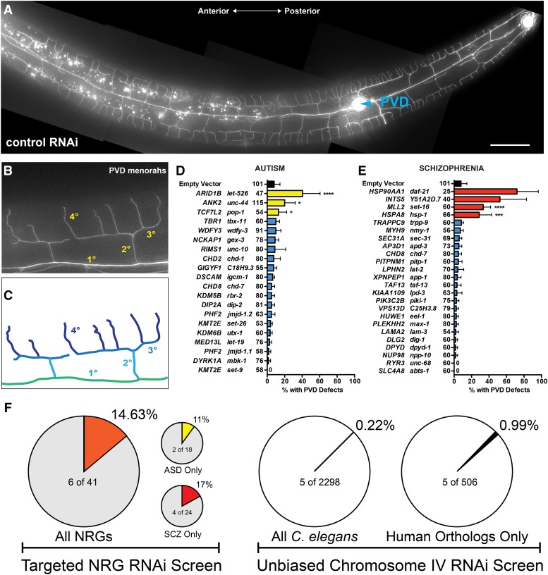
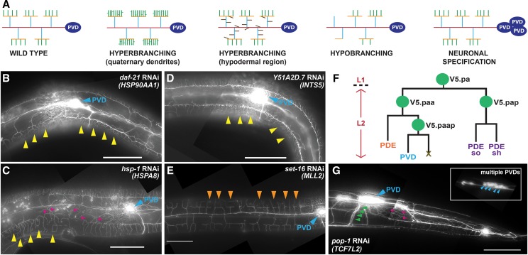
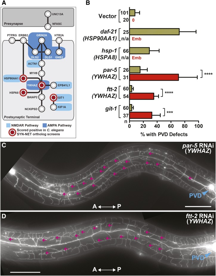
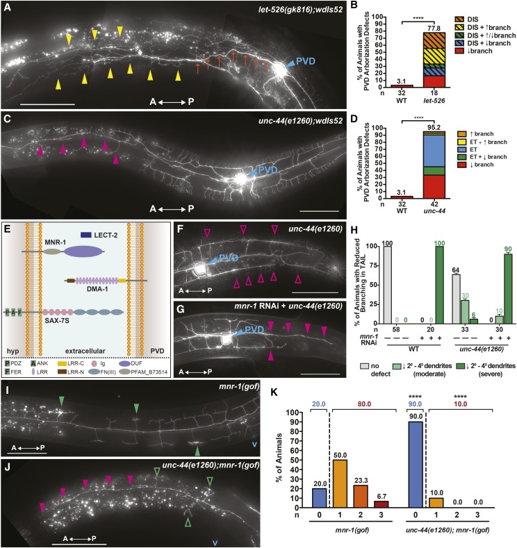
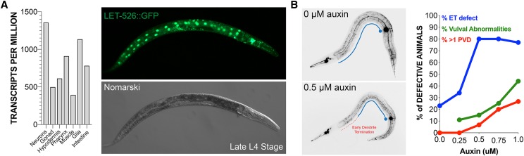
Analysis of patient-derived DNA samples has identified hundreds of variants that are likely involved in neuropsychiatric diseases such as autism spectrum disorder (ASD) and schizophrenia (SCZ). While these studies couple behavioral phenotypes to individual genotypes, the number and diversity of candidate genes implicated in these disorders highlights the fact that the mechanistic underpinnings of these disorders are largely unknown. Here, we describe a RNAi-based screening platform that uses C. elegans to screen candidate neuropsychiatric risk genes (NRGs) for roles in controlling dendritic arborization. To benchmark this approach, we queried published lists of NRGs whose variants in ASD and SCZ are predicted to result in complete or partial loss of gene function. We found that a significant fraction (>16%) of these candidate NRGs are essential for dendritic development. Furthermore, these gene sets are enriched for dendritic arbor phenotypes (>14 fold) when compared to control RNAi datasets of over 500 human orthologs. The diversity of PVD structural abnormalities observed in these assays suggests that the functions of diverse NRGs (encoding transcription factors, chromatin remodelers, molecular chaperones and cytoskeleton-related proteins) converge to regulate neuronal morphology and that individual NRGs may play distinct roles in dendritic branching. We also demonstrate that the experimental value of this platform by providing additional insights into the molecular frameworks of candidate NRGs. Specifically, we show that ANK2/UNC-44 function is directly integrated with known regulators of dendritic arborization and suggest that altering the dosage of ARID1B/LET-526 expression during development affects neuronal morphology without diminishing aspects of cell fate specification.

Keywords: Caenorhabditis elegans; RNA interference; autism spectrum disorder; dendritic arborization; model organism; neuronal development; neuropsychiatric risk genes; schizophrenia.


Figures

Figure 1
Figure 1
Proper Neuronal Development is Perturbed after the Depletion of C. elegans orthologs of ASD or SCZ NRGs. (A) PVD neurons form a web-like dendritic arbor that envelops the body of adult-staged animals. (B,C) PVD menorahs are individual dendritic units that sprout ventrally and dorsally from primary dendrites (1°). Each menorah is composed of secondary (2°), tertiary (3°), and quaternary (4°) dendrites. (D,E) RNAi knockdown of the C. elegans orthologs of (D) 3/18 ASD NRGs and (E) 4/24 SCZ NRGs resulted in PVD dendritic arborization or cell-fate specification defects. A single NRG, CHD8, was present in both the ASD- and SCZ-associated lists. Animals exhibiting an overt increase and/or decrease in dendritic branching, a disorganization of the dendritic arbor, or supernumerary PVD cell bodies were scored as defective. For each panel, human NRGs = left column and C. elegans orthologs of human NRGs = right column. (F) Comparison of the overall hit rates of ASD and SCZ candidates compared to unbiased screening. Error bars indicate the weighted standard deviation. ****P < 0.0001, ***P < 0.001, *P < 0.05 determined by Fisher’s exact test.
Figure 2
Figure 2
RNAi-mediated Gene Knockdown of NRG Orthologs Disrupts Dendritic Arbor Patterning and Neuronal Cell Fate Specification. (A) Schematic depicting the types of dendritic arborization or cell specification defects scored throughout all candidate-based RNAi screens in this study. RNAi against (B) daf-21 (HSP90AA1) or (D) Y51A2D.7 (INTS5) leads to hyperbranching of 4° dendrites (yellow arrowheads). (C) Animals fed hsp-1 (HSPA8) dsRNA exhibit hyperbranching of 4° dendrites (yellow arrowheads) and increased dendritic branching in the hypodermal region (magenta arrows). (E) RNAi against set-16 (MLL2) results in hypobranching of 4° dendrites (orange arrowheads). (F) Cell lineage diagram depicting stage-specific division patterns that give rise to PVD neurons. (G) pop-1 (TCF7L2) dsRNA-treated animals exhibit supernumerary PVD cell bodies (blue arrowheads, inset) and axons (green arrowheads) along with an increase in dendritic branching (magenta arrows). In panels B, C, D, E, and G, the PVD cell body is labeled with a blue arrowhead and anterior is to the left. Scale bars: 50μm.
Figure 3
Figure 3
Use of the RNAi Hypersensitive Strain, nre-1(hd20) lin-15b(hd126), offers a Complementary Approach to Identifying Additional NRG Orthologs that Regulate Dendritic Development. (A) A synaptic protein-protein interaction network in which SYN-NET NRG interactions were identified via the SynSysNet Database (http://bioinformatics.charite.de/synsysnet/). Circles demarcated in red are positive hits identified through the use of the wild-type or the nre-1(hd20) lin-15b(hd126) RNAi hypersensitive strain. (B) RNAi of all SYN-NET NRG orthologs identified four NRGs (represented by five C. elegans orthologs) as novel regulators of dendritic branching in either the wild-type (yellow bar with diagonal lines) or nre-1(hd20) lin-15b(hd126) RNAi hypersensitive (red bar) background. (C and D) YWHAZ orthologs, par-5 and ftt-2, exhibit increased dendritic branching in the hypodermal region (magenta arrows). Anteroposterior orientation is indicated by the white double-headed arrow. In panels d and e, the PVD cell body is labeled with a blue arrowhead. n, number of animals scored. Error bars indicate the weighted standard deviation. ****P < 0.0001, ***P < 0.001 determined by Fisher’s exact test. Scale bars: 50μm.
Figure 4
Figure 4
Genetic Mutants Phenocopy RNAi-induced Dendritic Arborization Defects and Genetic Interaction Studies Indicate that unc-44 (ANK2) is Required for SAX-7/MNR-1/LECT-2/DMA-1 Signaling. (A,B) let-526(gk816) genetic mutants primarily exhibit a marked disorganization of the dendritic arbor, including spurious branching (yellow arrowheads) and misguidance of the 1° dendrite (orange arrows). A general increase and/or decrease in dendritic branching may also accompany the disorganization phenotype. (C,D) In addition to early termination of the 1° anterior dendrite (not shown), unc-44(e1260) genetic mutants may also exhibit a loss of 2°, 3°, and 4° dendrites (magenta arrowheads) at the anterior end of the arbor. (E) Schematic depicting the SAX-7S/MNR-1/LECT-2/DMA-1 multi-protein signaling complex along with protein domains. (F) unc-44(e1260) genetic mutants largely retain the ability to form 4° dendrites (magenta open arrowheads) in the tail region, although 6% exhibit a complete reduction in 2°, 3°, and 4° dendrites (H). (G,H) A complete reduction in 2°, 3°, and 4° dendrites (magenta arrowheads) in the tail region is exhibited by 90% of unc-44(e1260) mutants treated with mnr-1 dsRNA. (I,-K) 80% of mnr-1(gof) animals elaborate ≥ 1 baobab (green arrowheads) anterior to the vulva, while 10% of unc-44(e1260);mnr-1(gof) animals sprout ≥ 1 baobab (green open arrowheads) in this region. mnr-1(gof) animals harboring the unc-44(e1260) allele also exhibit a loss of 2°, 3°, and 4° dendrites (magenta arrowheads) at the anterior end of the PVD arbor. Anteroposterior orientation is indicated by the white, double-headed arrow. In panels A, C, F, and G, the PVD cell body is labeled with a blue arrowhead, and “v” in panels i and j marks the approximate location of the vulva. In panels b, d, and h, “n” is the number of animals scored. In panel K, “n” is the number of baobabs present in each genetic background. ****P < 0.0001 determined by Fisher’s exact test. Scale bars: 50μm.
Figure 5
Figure 5
Dendritic architecture is extremely sensitive to reductions in LET-526/ARID1B expression. (A) Single cell RNA-seq experiments (Cao et al. 2017) indicate that let-526 mRNAs are highly expressed in all major tissue types in developing animals. Consistent these results, LET-526::GFP is also broadly expressed. (B) Animals were treated with a gradation of auxin during development. Relatively low concentrations of auxin exposure to animals harboring a let-526::degron allele resulted in dendritic arborization phenotypes that mirrored those found in let-536(lf) mutant animals. The neuronal morphology defects during PVD development were elicited at much lower concentrations of auxin that are needed to observe other pleiotropic somatic phenotypes. ET= extension defects.

References

    1. Aguirre-Chen C., Bülow H. E., and Kaprielian Z., 2011. C. elegans bicd-1, homolog of the Drosophila dynein accessory factor Bicaudal D, regulates the branching of PVD sensory neuron dendrites. Development 138: 507–518. 10.1242/dev.060939 - DOI - PMC - PubMed
    1. Albeg A., Smith C. J., Chatzigeorgiou M., Feitelson D. G., Hall D. H. et al. , 2011. C. elegans multi-dendritic sensory neurons: morphology and function. Mol. Cell. Neurosci. 46: 308–317. 10.1016/j.mcn.2010.10.001 - DOI - PMC - PubMed
    1. An J. Y., Cristino A. S., Zhao Q., Edson J., Williams S. M. et al. , 2014. Towards a molecular characterization of autism spectrum disorders: an exome sequencing and systems approach. Transl. Psychiatry 4: e394 10.1038/tp.2014.38 - DOI - PMC - PubMed
    1. Bhattacharya A., Aghayeva U., Berghoff E. G., and Hobert O., 2019. Plasticity of the Electrical Connectome of C. elegans. Cell 176: 1174–1189.e16. 10.1016/j.cell.2018.12.024 - DOI - PMC - PubMed
    1. Blake J. A., Bult C. J., Kadin J. A., Richardson J. E., and Eppig J. T., Mouse Genome Database Group , 2011. The Mouse Genome Database (MGD): premier model organism resource for mammalian genomics and genetics. Nucleic Acids Res. 39: D842–D848. 10.1093/nar/gkq1008 - DOI - PMC - PubMed

Publication types

MeSH terms

Substances