

Fluorescent Hybridization of Mycobacterium leprae in Skin Samples Collected in Burkina Faso
- PMID: 32132193
- PMCID: PMC7180235
- DOI: 10.1128/JCM.02130-19
Fluorescent Hybridization of Mycobacterium leprae in Skin Samples Collected in Burkina Faso
Abstract
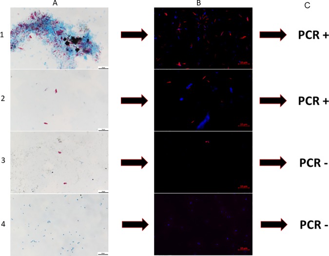
Leprosy is caused by Mycobacterium leprae, and it remains underdiagnosed in Burkina Faso. We investigated the use of fluorescent in situ hybridization (FISH) for detecting M. leprae in 27 skin samples (skin biopsy samples, slit skin samples, and skin lesion swabs) collected from 21 patients from Burkina Faso and three from Côte d'Ivoire who were suspected of having cutaneous leprosy. In all seven Ziehl-Neelsen-positive skin samples (four skin biopsy samples and three skin swabs collected from the same patient), FISH specifically identified M. leprae, including one FISH-positive skin biopsy sample that remained negative after testing with PCR targeting the rpoB gene and with the GenoType LepraeDR assay. Twenty other skin samples and three negative controls all remained negative for Ziehl-Neelsen staining, FISH, and rpoB PCR. These data indicate the usefulness of a microscopic examination of skin samples after FISH for first-line diagnosis of cutaneous leprosy. Accordingly, FISH represents a potentially useful point-of-care test for the diagnosis of cutaneous leprosy.
Keywords: Mycobacterium; Mycobacterium leprae; fluorescent hybridization; leprosy; skin.
Copyright © 2020 American Society for Microbiology.
Figures
References
-
- Ouédraogo N, Ouédraogo M, Tapsoba G, Traore FF, Bassole A, Zeba/Lompo S, Tioye YL, Ilboudo L, Kabore N, Zigani E, Drabo F, Serme M, Nassa C, Kafando C, Korsaga/Some NN, Barro/Traore F, Niamba P, Traore A. 2018. Dépistage de la lèpre en stratégie avancée au Burkina Faso. Bull Assoc Lepr Langue Fr 33:4–7. https://www.leprosy-information.org/media/838/download.
-
- Shepard CC, McRae DH. 1968. A method for counting acid-fast bacteria. Int J Lepr Other Mycobact Dis 36:78–82. - PubMed
Publication types
MeSH terms
Substances
LinkOut - more resources
Full Text Sources
Medical
