. 2020 Mar 17;117(11):5883-5894.
doi: 10.1073/pnas.1912055117. Epub 2020 Mar 4.

TDP-43 α-helical structure tunes liquid-liquid phase separation and function

Affiliations

TDP-43 α-helical structure tunes liquid-liquid phase separation and function

Alexander E Conicella et al. Proc Natl Acad Sci U S A. .

Abstract

Liquid-liquid phase separation (LLPS) is involved in the formation of membraneless organelles (MLOs) associated with RNA processing. The RNA-binding protein TDP-43 is present in several MLOs, undergoes LLPS, and has been linked to the pathogenesis of amyotrophic lateral sclerosis (ALS). While some ALS-associated mutations in TDP-43 disrupt self-interaction and function, here we show that designed single mutations can enhance TDP-43 assembly and function via modulating helical structure. Using molecular simulation and NMR spectroscopy, we observe large structural changes upon dimerization of TDP-43. Two conserved glycine residues (G335 and G338) are potent inhibitors of helical extension and helix-helix interaction, which are removed in part by variants at these positions, including the ALS-associated G335D. Substitution to helix-enhancing alanine at either of these positions dramatically enhances phase separation in vitro and decreases fluidity of phase-separated TDP-43 reporter compartments in cells. Furthermore, G335A increases TDP-43 splicing function in a minigene assay. Therefore, the TDP-43 helical region serves as a short but uniquely tunable module where application of biophysical principles can precisely control assembly and function in cellular and synthetic biology applications of LLPS.

Keywords: NMR spectroscopy; liquid–liquid phase separation; molecular simulation; protein interactions.


Conflict of interest statement

The authors declare no competing interest.

Figures
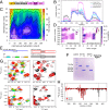
Fig. 1.
Fig. 1.
TDP-43 CTD self-associates and forms transient helical structures. (A) Domain structure of TDP-43. (B) α-Helical content of TDP-43 simulations at each residue, where single chain comes from a separate simulation of a single TDP-43310–350 chain (single chain, black), and the other three curves from a two-chain simulation using all frames (two chain [all], cyan), only strongly bound frames (strongly bound, magenta), or only the weakly bound frames (weakly bound, blue). Data are plotted as mean ± SEM of n = 5 equal divisions of the total dataset. (C) Free energy landscape of TDP-43310–350 intermolecular contacts for all-atom two-chain simulations highlight two minima corresponding to strongly and weakly bound states. (D) α-Helical secondary-structure propensity maps show the contribution of all helical configurations to the overall helical content shown in B. This analysis highlights stabilization of contiguous α-helical structure in the strongly bound state. (E) Schematic of cross-linked CTD variants. (F) Nonreducing SDS/PAGE of dimeric S273C and S387C illustrate an apparent reduction of molecular weight by approximately one-half upon addition of disulfide-breaking reducing agent (1 mM DTT). (G, Left) Select regions from 1H–15N TROSY spectra of dilsulfide-linked homodimeric TDP-43 CTD S273C (red) show line broadening and large upfield 15N chemical shift differences in 321–343 region compared to monomeric reduced S273C (blue), consistent with formation of structure. Chemical shifts for residues in the dimer state were assigned by extrapolation (dashed black line) from spectra of 20 µM (cyan), 45 µM (green), and 90 µM (orange) WT. (G, Right) Overlay of 1H–15N TROSY spectra regions for homodimeric S273C and S387C are similar in the central 320–340 region. (H) Quantification of 15N ∆δ values between homodimeric S273C (red) and S387C (black) and their respective monomers. The gray boxes indicate the positions of S273C and S387C disulfide cross-links.
Fig. 2.
Fig. 2.
G335 and G338 substitutions enhance TDP-43 CTD helical stability. (A) Per-residue α-helical propensities for WT, G335A, and G338A for atomistic single-chain simulations of TDP-43310–350 show higher fraction of α-helical structure near the sites of mutation (highlighted by red, G335A, or orange, G338A, bars). Data are plotted as mean ± SEM of n = 5 equal divisions of the total dataset. (B) Average simulated helicity is mostly unaffected for residues 321–330 (Left) away from G335 and G338 mutation sites but enhanced for 331–343 (Right) surrounding the mutation site. (C) Maps of contiguous α-helical structure location (y axis) and length (x axis) for TDP-43310–350 show increased probability for longer helix structure in G335A and G338A relative to WT. One example configuration from a subpopulation of the simulation ensemble (indicated by the black arrow) is displayed for each variant. (D) Experimental NMR secondary chemical shifts (ΔδCαΔδCβ) and the differences in secondary shifts with respect to WT (∆∆δ) for G335A and G338A show increased α-helical structure near the site of mutation. The secondary shift values for WT are overlaid in black for comparison. (E) The average experimental secondary shift (ΔδCαΔδCβ¯) for TDP-43 331–343 of WT and mutant CTD highlight increases in local α-helical structure in all variants. Error bars are SEM. (F) Higher {1H}–15N heteronuclear nuclear Overhauser effect (NOE) values measured for G335A (red) and G338A (orange) compared to WT (black) near the site of mutation indicate slowed local protein backbone motion, consistent with enhanced helical structure. Mutation positions G335A and G338A highlighted with red and orange lines, respectively. For all above panels: Simulation data are plotted as mean and SEM from 10 equal blocks of equilibrated structural ensemble. Unless otherwise stated, experimental data are plotted as mean and SD estimated from signal-to-noise ratio.
Fig. 3.
Fig. 3.
Mutations enhance TDP-43 CTD helix–helix interaction and assembly. (A) Larger chemical shift differences (∆δ) in the helical region of TDP-43 CTD for G335A and G338A compared to WT are consistent with enhanced helix–helix interactions. Shifts are reported at concentrations from 20 to 90 µM with respect to a monomeric (low concentration) reference (20 µM for WT and G335A; 10 µM for G338A). The gray boxes indicate the loss of detectable peak intensity due to line broadening. (B) Normalized chemical shift differences vs. concentration are higher for mutant CTD compared to WT, consistent with increased self-assembly for G335 and G338 variants. Error bars are SD estimated by 100 bootstrapping simulations derived from experimental uncertainty. (C) Diffusion coefficients calculated from NMR diffusion data plotted as a function of CTD concentration show that WT (black) and G335A (red) diffuse slower than A326P (green). Average diffusion coefficients for S273C and S387C dimers and S273C and S387C monomers are indicated by blue and black horizontal lines, respectively. Data are plotted as mean ± SD, indicated for monomer and dimer diffusion coefficients by gray and light blue boxes, respectively.
Fig. 4.
Fig. 4.
Mutations at G335 and G338 alter in vitro LLPS. (A) Protein remaining in supernatant after phase separation for WT and mutant CTD measured for 0 to 500 mM NaCl. Error bars represent SD of three replicates. (B) DIC micrographs of LLPS for WT TDP-43 CTD and variants G335A, G335D, G335N, G335S, and G338A at 0 and 60 min after addition of NaCl to 300 mM. (Scale bars, 20 µm.) (C) The protein concentration remaining in supernatant of WT and mutant CTD at 150 mM NaCl (“physiological conditions”) correlates well with the average change in secondary shift (Δδ¯) (Pearson r = −0.95). Error bars represent SD of concentrations using three replicates and SEM for ΔδCαΔδCβ¯. (D) Coexistence curves of TDP-43 CTD from coarse-grained (CG) slab simulations of WT and G335/G338 mutants illustrate no discernable difference in LLPS behavior as a result of mutation. The Inset shows the low-concentration phase on a log scale and highlights the similarity even at very low concentrations. (E) Coexistence curves of TDP-43 from CG slab simulations illustrate enhanced phase separation when residues 320–334 are fixed as a rigid helix (magenta) instead of fully flexible (black). Inset shows the approximately twofold change to left arm of the phase diagram with temperature vs. concentration on a log scale. (F) A snapshot of the TDP-43 CTD CG slab simulation where CTD molecules harboring rigid α-helical substructure (magenta) visit both dispersed and condensed states in phase coexistence. Arrows indicate oligomers formed by helix–helix contacts.
Fig. 5.
Fig. 5.
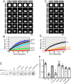
Mutations at G335 and G338 alter in-cell LLPS fluidity and splicing function. (AD) Fluorescence recovery after in-cell half-droplet photobleaching and quantification for TDP-43RRM-GFP reporter construct with (A and B) WT, G335D, G335N, G335S, and G335A, or (C and D) WT, G335A, and G338A CTD sequences. TDP-43RRM-GFP shows differential droplet fluidity for G335 variants relative to WT. (E) Example agarose gel of PCR products to quantify the fraction of exon 9 CFTR exclusion (OUT) expressed from a minigene construct transfected in HeLa cells. Cells were treated with control, TDP-43 siRNA, or TDP-43 siRNA plus WT or mutant vectors. (F) The splicing activity was calculated as the ratio of exon 9 exclusion in HeLa cells expressing siRNA-resistant WT and mutant constructs (as labeled) or in absence of exogenous TDP-43 expression (T2). Nonspecific siRNA was used as control (N). Error bars are SEM of three or more replicates.

References

    1. Kato M., et al. , Cell-free formation of RNA granules: Low complexity sequence domains form dynamic fibers within hydrogels. Cell 149, 753–767 (2012). - PMC - PubMed
    1. Mitrea D. M., et al. , Nucleophosmin integrates within the nucleolus via multi-modal interactions with proteins displaying R-rich linear motifs and rRNA. eLife 5, 1–33 (2016). - PMC - PubMed
    1. Nott T. J., et al. , Phase transition of a disordered nuage protein generates environmentally responsive membraneless organelles. Mol. Cell 57, 936–947 (2015). - PMC - PubMed
    1. Brangwynne C., Hoege C., Gharakhani J., Jülicher F., Hyman A. A., Germline P granules are liquid droplets that localize by controlled dissolution/condensation. Science 324, 1729–1732 (2009). - PubMed
    1. Hyman A. A., Weber C. A., Jülicher F., Liquid-liquid phase separation in biology. Annu. Rev. Cell Dev. Biol. 30, 39–58 (2014). - PubMed

Publication types

MeSH terms