. 2020 Mar 17;117(11):6196-6204.
doi: 10.1073/pnas.1919303117. Epub 2020 Mar 4.

Anterior cingulate cortex is necessary for adaptation of action plans

Affiliations

Anterior cingulate cortex is necessary for adaptation of action plans

Adam T Brockett et al. Proc Natl Acad Sci U S A. .

Abstract

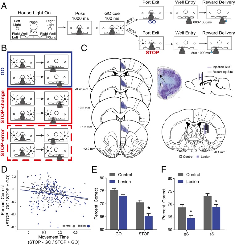
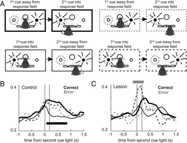
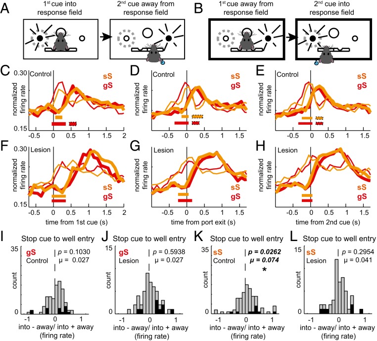
Previous research has focused on the anterior cingulate cortex (ACC) as a key brain region in the mitigation of the competition that arises from two simultaneously active signals. However, to date, no study has demonstrated that ACC is necessary for this form of behavioral flexibility, nor have any studies shown that ACC acts by modulating downstream brain regions such as the dorsal medial striatum (DMS) that encode action plans necessary for task completion. Here, we performed unilateral excitotoxic lesions of ACC while recording downstream from the ipsilateral hemisphere of DMS in rats, performing a variant of the STOP-signal task. We show that on STOP trials lesioned rats perform worse, in part due to the failure of timely directional action plans to emerge in the DMS, as well as the overrepresentation of the to-be-inhibited behavior. Collectively, our findings suggest that ACC is necessary for the mitigation of competing inputs and validates many of the existing theoretical predictions for the role of ACC in cognitive control.

Keywords: action planning; anterior cingulate; conflict; inhibition; striatum.


Conflict of interest statement

The authors declare no competing interest.

Figures

Fig. 1.
Fig. 1.
Unilateral ACC lesions impair response selection. (A) Illustration of STOP-change task. Rats were required to nose-poke and remain in the port for 1,000 ms before one of two directional lights (left or right) were flashed for 100 ms. For 80% of the time, the cue light directs the rat toward the correct fluid well (GO trials). For 20% of the time, following port withdrawal (0 to 100 ms), the cue light opposite the first cue light turns on and remains on, instructing the rat to inhibit its response to the first cue light in favor of responding to the second cue light (STOP-change trials). (B) Illustration of GO (blue), STOP-change correct (red), and STOP-change error (dotted red) trials. (C, Left) Stereotaxic overlays detailing the extent of the lesion for all eight rats (four females, four males) tested. Lesions were primarily constrained to rodent Cg1 and showed little overlap with other frontal regions (35). (C, Middle) Photomicrograph of Nissl-stained tissue showing representative lesion (arrow). Overlay approximately matched to section (∼+1.6 mm anterior from bregma). (Scale bar, 1 mm.) (C, Top Right) Sagittal view of two injection sites in ACC and the targeted recording site in DMS. (C, Bottom Right) Overlays detailing the tracks of all 16 electrode tracks in the DMS. Shaded blue tracks signify lesioned animals, while black borders signify controls. Injections and electrodes were placed in either the right or the left hemisphere, and placement was counterbalanced across treatment groups. Note: placements are shown on the same hemisphere to reflect the unilateral nature of the manipulations. (D) Scatter plot showing the percentage of correct responses as a function of movement time. Black and blue dots indicate data from individual sessions for controls and lesions, respectively. (E) Percentage of correct scores on GO and STOP-change trials by condition averaged over recording sessions. (F) Percentage of correct scores based on sequence effects averaged over recording sessions. Lowercase letters indicate previous trial type; uppercase letters indicate current trial type (gS reads as “GO trial preceded a STOP-change trial”). A single asterisk (*) indicates significance at Bonferroni-adjusted P < 0.05.
Fig. 2.
Fig. 2.
Unilateral ACC lesions delay response selection in the DMS on STOP-change trials. (A) Illustration of trial types as they relate to movement into and away from a neuron’s response field for GO (blue) and STOP (red) trials as defined by firing in response to the first cue light. (B and E) Population histogram for control (B; n = 97) and lesioned (E; n = 53) rats aligned to port exit. Blue lines represent GO trials; red lines represent correct STOP-change trials. Thin lines represent movements away from a neuron’s response field (nonpreferred direction); thick lines represent movement into a neuron’s response field (preferred direction). Preferred direction was defined by the direction that elicited the strongest response averaged over correct trial types. Vertical dashed line represents the SCRT as computed by subtracting movement times on GO trials from STOP trials. (C and F) Directional indices (into − away/into + away) for firing related to the first cue-light epoch (first cue-light onset to port exit) in controls (C) and lesioned (F) rats. (D and G) Directional indices for firing related to the second cue-light epoch (second cue light to well entry) in controls (D) and lesioned (G) rats. A single asterisk (*) indicates significance at Wilcoxon P < 0.05.
Fig. 3.
Fig. 3.
Firing in DMS is overly active during errant responses after ACC lesions. (A) Illustration of behavioral responses on STOP-change correct (solid) and STOP-change error (dashed) trials. (B and C) Population histograms for control (B; n = 97) and lesion (C; n = 53) rats aligned to onset of the second cue light. Solid lines represent firing on STOP-change trials where the rat made the correct response, and dashed lines represent STOP-change trials where the rat committed an error. Thick and thin lines represent firing into (thick) or away from (thin) a neuron’s preferred direction. Tick marks (gray) represent significance when comparing thick to thin lines within trial type.
Fig. 4.
Fig. 4.
Unilateral ACC lesions disrupt STOP-change trial performance regardless of previous experience. (A and B) Illustrations of responding into and away from a neuron’s response field. (C and F) Population histograms comparing sequence effects in control (C; n = 89) and lesioned (F; n = 48) rats. Red lines represent gS trials, and orange lines represent sS trials. Thin lines represent movement away from the response field. Thick lines represent movement into the response field. The histogram is aligned to the first cue. Tick marks represent significance when comparing thick to thin lines within a trial type. (D and G) Population histograms comparing sequence effects between control (D) and lesioned (G) rats when aligned to port exit. (E and H) Population histograms comparing sequence effects between control (E) and lesioned (H) rats when aligned to the second cue. (IL) Distributions of directional indices (into − away/into + away) during the second cue-light epoch (stop cue to well entry) for gS (I and J) and sS trials (K and L) for control (I and K) and lesioned (J and L) rats. The single asterisk (*) indicates significance at Wilcoxon P < 0.05.

References

    1. Verbruggen F., et al. , A consensus guide to capturing the ability to inhibit actions and impulsive behaviors in the stop-signal task. eLife 8, e46323 (2019). - PMC - PubMed
    1. Shenhav A., Cohen J. D., Botvinick M. M., Dorsal anterior cingulate cortex and the value of control. Nat. Neurosci. 19, 1286–1291 (2016). - PubMed
    1. Ebitz R. B., Hayden B. Y., Dorsal anterior cingulate: A Rorschach test for cognitive neuroscience. Nat. Neurosci. 19, 1278–1279 (2016). - PubMed
    1. Kolling N., et al. , Value, search, persistence and model updating in anterior cingulate cortex. Nat. Neurosci. 19, 1280–1285 (2016). - PMC - PubMed
    1. Stolyarova A., et al. , Dissociable roles for anterior cingulate cortex and basolateral amygdala in decision confidence and learning under uncertainty. bioRxiv, 10.1101/655860 (2019). - PMC - PubMed

Publication types

LinkOut - more resources