

Anatomy and function of the fornix in the context of its potential as a therapeutic target
- PMID: 32132227
- PMCID: PMC7231447
- DOI: 10.1136/jnnp-2019-322375
Anatomy and function of the fornix in the context of its potential as a therapeutic target
Abstract
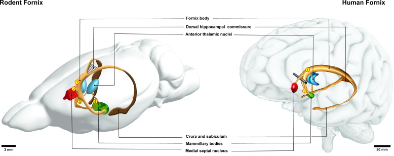
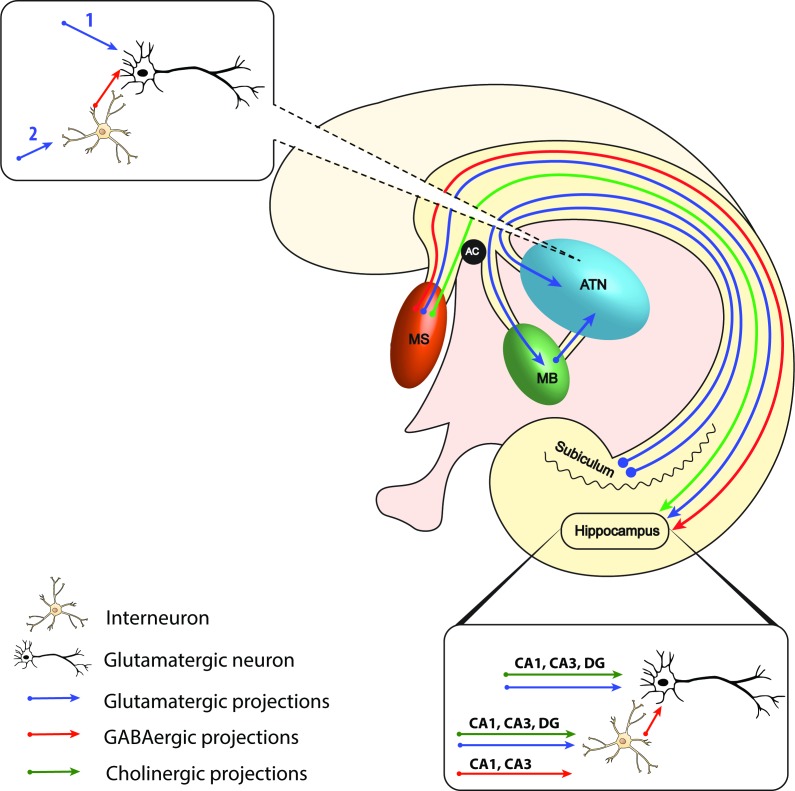
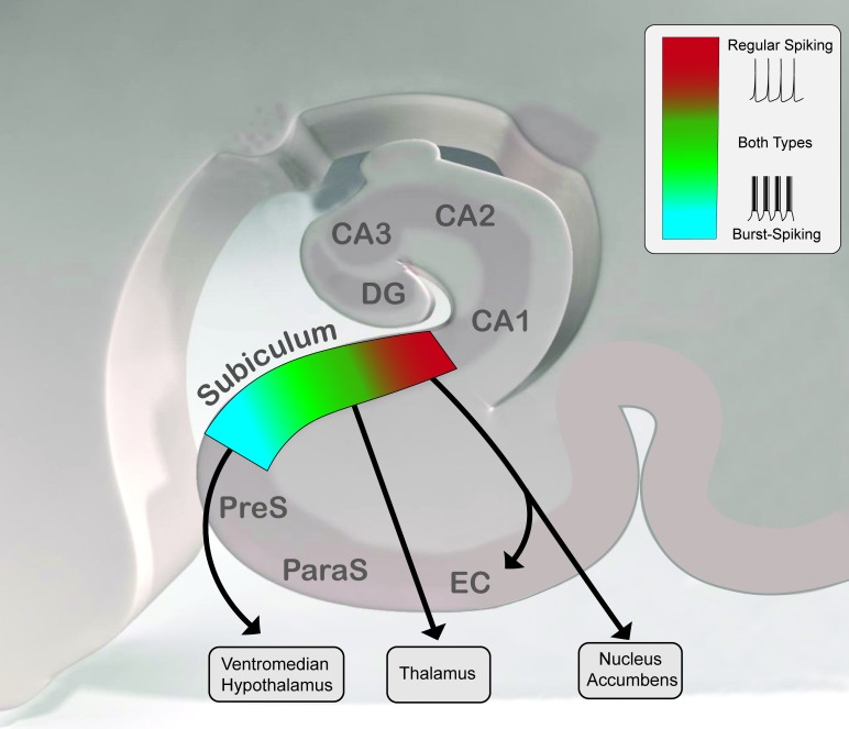
The fornix is a white matter bundle located in the mesial aspect of the cerebral hemispheres, which connects various nodes of a limbic circuitry and is believed to play a key role in cognition and episodic memory recall. As the most prevalent cause of dementia, Alzheimer's disease (AD) dramatically impairs the quality of life of patients and imposes a significant societal burden on the healthcare system. As an established treatment for movement disorders, deep brain stimulation (DBS) is currently being investigated in preclinical and clinical studies for treatment of memory impairment in AD by modulating fornix activity. Optimal target and stimulation parameters to potentially rescue memory deficits have yet to be determined. The aim of this review is to consolidate the structural and functional aspects of the fornix in the context of neuromodulation for memory deficits. We first present an anatomical and functional overview of the fibres and structures interconnected by the fornix. Recent evidence from preclinical models suggests that the fornix is subdivided into two distinct functional axes: a septohippocampal pathway and a subiculothalamic pathway. Each pathway's target and origin structures are presented, followed by a discussion of their oscillatory dynamics and functional connectivity. Overall, neuromodulation of each pathway of the fornix is discussed in the context of evidence-based forniceal DBS strategies. It is not yet known whether driving fornix activity can enhance cognition-optimal target and stimulation parameters to rescue memory deficits have yet to be determined.
Keywords: alzheimer's disease; electrical stimulation; limbic system; neurosurgery.
© Author(s) (or their employer(s)) 2020. Re-use permitted under CC BY-NC. No commercial re-use. See rights and permissions. Published by BMJ.
Conflict of interest statement
Competing interests: AML is a consultant to Medtronic, St Jude, Boston Scientific, Functional Neuromodulation and Insightec.
Figures
Similar articles
-
Deep brain stimulation of fornix in Alzheimer's disease: From basic research to clinical practice.Eur J Clin Invest. 2023 Aug;53(8):e13995. doi: 10.1111/eci.13995. Epub 2023 Apr 10. Eur J Clin Invest. 2023. PMID: 37004153 Review.
-
Utilizing diffusion tensor imaging as an image biomarker in exploring the therapeutic efficacy of forniceal deep brain stimulation in a mice model of Alzheimer's disease.J Neural Eng. 2024 Sep 4;21(5). doi: 10.1088/1741-2552/ad7322. J Neural Eng. 2024. PMID: 39230033
-
The effect of fornix deep brain stimulation in brain diseases.Cell Mol Life Sci. 2020 Sep;77(17):3279-3291. doi: 10.1007/s00018-020-03456-4. Epub 2020 Jan 23. Cell Mol Life Sci. 2020. PMID: 31974655 Free PMC article. Review.
-
Fornical Closed-Loop Stimulation for Alzheimer's Disease.Trends Neurosci. 2018 Jul;41(7):418-428. doi: 10.1016/j.tins.2018.03.015. Epub 2018 May 4. Trends Neurosci. 2018. PMID: 29735372 Review.
-
Neuronal Network Activation Induced by Forniceal Deep Brain Stimulation in Mice.Genes (Basel). 2025 Feb 9;16(2):210. doi: 10.3390/genes16020210. Genes (Basel). 2025. PMID: 40004540 Free PMC article.
Cited by
-
Behavior is movement only but how to interpret it? Problems and pitfalls in translational neuroscience-a 40-year experience.Front Behav Neurosci. 2022 Oct 5;16:958067. doi: 10.3389/fnbeh.2022.958067. eCollection 2022. Front Behav Neurosci. 2022. PMID: 36330050 Free PMC article. Review.
-
Structural Alterations of the Corpus Callosum in Children With Infantile Hydrocephalus.J Child Neurol. 2024 Jan;39(1-2):66-76. doi: 10.1177/08830738241231343. Epub 2024 Feb 22. J Child Neurol. 2024. PMID: 38387869 Free PMC article.
-
Deriving causal relationships in resting-state functional connectivity using SSFO-based optogenetic fMRI.J Neural Eng. 2022 Nov 8;19(6):10.1088/1741-2552/ac9d66. doi: 10.1088/1741-2552/ac9d66. J Neural Eng. 2022. PMID: 36301683 Free PMC article.
-
Choir singing is associated with enhanced structural connectivity across the adult lifespan.Hum Brain Mapp. 2024 May;45(7):e26705. doi: 10.1002/hbm.26705. Hum Brain Mapp. 2024. PMID: 38716698 Free PMC article.
-
Diffusion tensor imaging biomarkers for assessing cognitive and physical function in aging.J Neuroeng Rehabil. 2025 Jul 10;22(1):157. doi: 10.1186/s12984-025-01698-6. J Neuroeng Rehabil. 2025. PMID: 40640846 Free PMC article.
References
-
- ADvance II Study: DBS-f in Patients With Mild Alzheimer’s Disease. ClinicalTrials.gov 2019.
Publication types
MeSH terms
LinkOut - more resources
Full Text Sources
Research Materials