. 2020 May 4;94(10):e01945-19.
doi: 10.1128/JVI.01945-19. Print 2020 May 4.

The Transcriptional Cofactor VGLL1 Drives Transcription of Human Papillomavirus Early Genes via TEAD1

Affiliations

The Transcriptional Cofactor VGLL1 Drives Transcription of Human Papillomavirus Early Genes via TEAD1

Seiichiro Mori et al. J Virol. .

Abstract

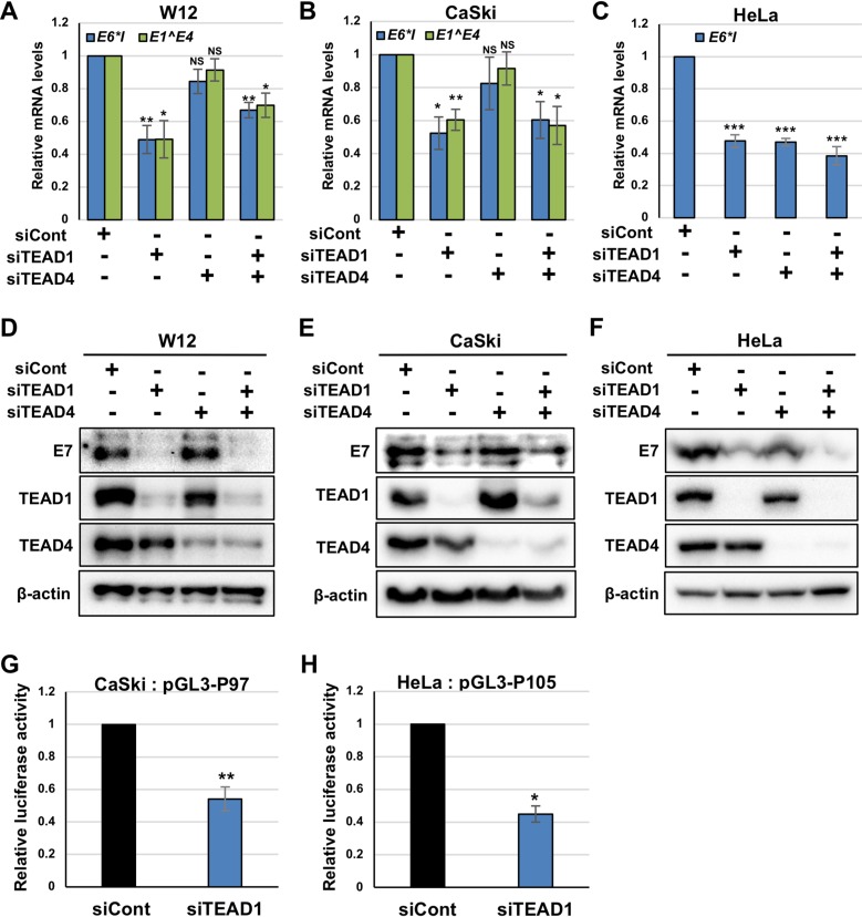
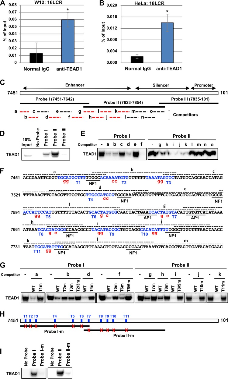
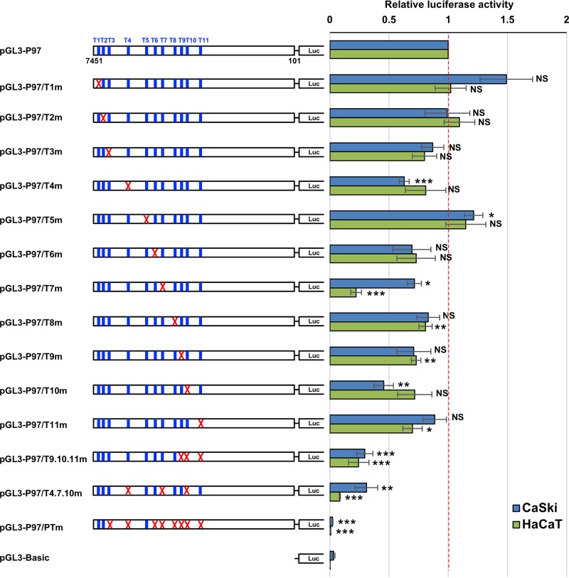
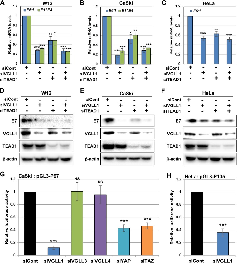
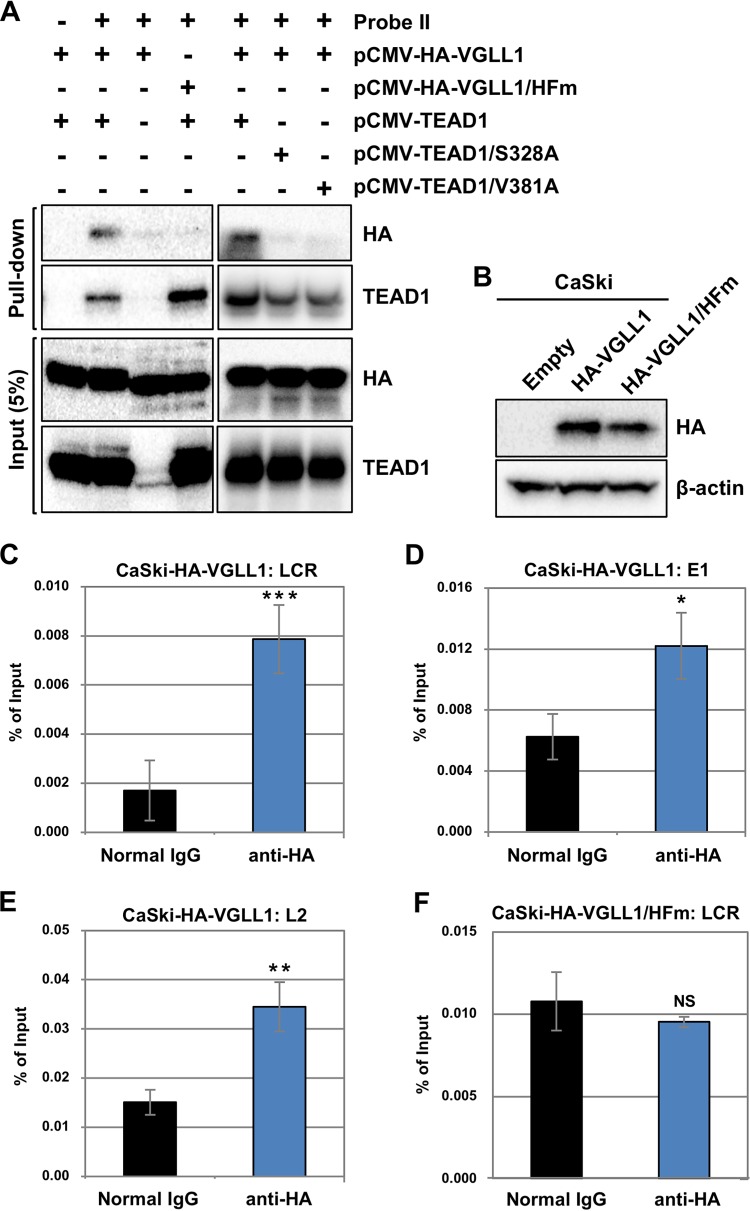
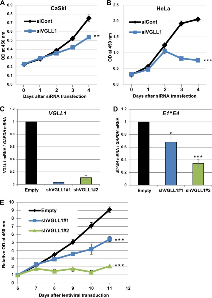
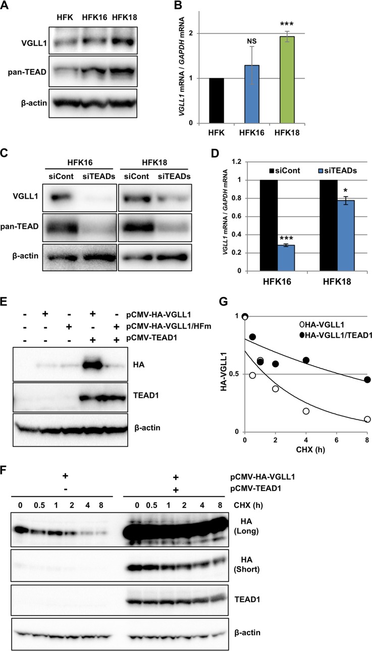
The TEAD family of transcription factors requires associating cofactors to induce gene expression. TEAD1 is known to activate the early promoter of human papillomavirus (HPV), but the precise mechanisms of TEAD1-mediated transactivation of the HPV promoter, including its relevant cofactors, remain unexplored. Here, we reveal that VGLL1, a TEAD-interacting cofactor, contributes to HPV early gene expression. Knockdown of VGLL1 and/or TEAD1 led to a decrease in viral early gene expression in human cervical keratinocytes and cervical cancer cell lines. We identified 11 TEAD1 target sites in the HPV16 long control region (LCR) by in vitro DNA pulldown assays; 8 of these sites contributed to the transcriptional activation of the early promoter in luciferase reporter assays. VGLL1 bound to the HPV16 LCR via its interaction with TEAD1 both in vitro and in vivo Furthermore, introducing HPV16 and HPV18 whole genomes into primary human keratinocytes led to increased levels of VGLL1, due in part to the upregulation of TEADs. These results suggest that multiple VGLL1/TEAD1 complexes are recruited to the LCR to support the efficient transcription of HPV early genes.IMPORTANCE Although a number of transcription factors have been reported to be involved in HPV gene expression, little is known about the cofactors that support HPV transcription. In this study, we demonstrate that the transcriptional cofactor VGLL1 plays a prominent role in HPV early gene expression, dependent on its association with the transcription factor TEAD1. Whereas TEAD1 is ubiquitously expressed in a variety of tissues, VGLL1 displays tissue-specific expression and is implicated in the development and differentiation of epithelial lineage tissues, where HPV gene expression occurs. Our results suggest that VGLL1 may contribute to the epithelial specificity of HPV gene expression, providing new insights into the mechanisms that regulate HPV infection. Further, VGLL1 is also critical for the growth of cervical cancer cells and may represent a novel therapeutic target for HPV-associated cancers.

Keywords: HPV; gene expression; transcription cofactor; transcription factor.


Figures

FIG 1
FIG 1
TEAD1 and TEAD4 regulate HPV early gene expression. (A to F) W12 (A and D), CaSki (B and E), and HeLa (C and F) cells were transfected with the indicated siRNA. At 2 days after transfection, the levels of HPV16 E6*I and E1̂E4 mRNAs (A and B) and HPV18 E6*I mRNA (C) were quantified by RT-qPCR and normalized to the level of GAPDH mRNA. The HPV16 E7 (D and E) and HPV18 E7 (F) proteins were detected by immunoblotting with anti-HPV16 and anti-HPV18 E7 antibodies, respectively. The effects of siRNA were verified by immunoblotting with anti-TEAD1 and anti-TEAD4 antibodies. β-Actin was used as the loading control. (G and H) CaSki (G) and HeLa (H) cells were transfected with the indicated siRNAs. Six hours later, the transfected CaSki and HeLa cells were further transfected with pGL3-P97 and pGL3-P105, respectively, together with the Renilla luciferase plasmid. At 2 days after transfection, firefly luciferase activity was measured and normalized to the Renilla luciferase activity after background subtraction. The quantitative data are the averages from three independent experiments, with the error bars representing the standard deviations. P values were determined by Student’s t test. NS, not significant (P > 0.05); *, P < 0.05; **, P < 0.01; ***, P < 0.005.
FIG 2
FIG 2
TEAD1 binds to the HPV LCRs. (A and B) Cross-linked chromatin prepared from W12 (A) and HeLa (B) cells was immunoprecipitated with an anti-TEAD1 antibody or normal rabbit IgG, and the recovered DNA was quantified by real-time PCR with primers for the HPV16 (A) and HPV18 (B) LCRs, respectively (16LCR and 18LCR, respectively). The levels of TEAD1 binding to the HPV16 or HPV18 genome are shown as percentages of the amount of input DNA. The data are averages from three experiments performed using independent chromatin preparations, with the error bars representing standard deviations. *, P < 0.05 (Student’s t test). (C) Schematic representation of the HPV16 LCR (nt 7451 to 101). The enhancer, silencer, and promoter regions are defined according to a previous study (52). The biotinylated DNA probes (probes I, II, and III) used in the DNA pulldown assays are indicated by solid lines. Oligonucleotide competitors (competitors a to o) are indicated by dashed lines. The competitors indicated by the red dashed lines inhibited TEAD1 binding to the probes (data shown in panel E). (D) The indicated biotinylated DNA probes were coupled to Dynabeads/M-280 streptavidin and incubated with HeLa nuclear extract; 10% of the input volume and the entire precipitated fractions were analyzed by immunoblotting using anti-TEAD1 antibody. (E) Unlabeled oligonucleotide competitors (a to o) were added to the binding reaction mixture, and the competition of TEAD1 binding against probe I or II was examined by DNA pulldown assays, as described in the legend to panel D. (F) Nucleotide sequence spanning the region from nt 7451 to 7800 of the HPV16 LCR. The TEAD-binding motifs (T1 to T11) are indicated in blue. The regions corresponding to the competitors (competitors a to m) are indicated by dashed lines over the nucleotide sequence. The nucleotide sequences in the mutant competitors and probes are denoted in red. The previously identified binding motifs for NF1 and AP1 are underlined. (G) Unlabeled oligonucleotide competitors or those having mutations were added to the binding reaction mixtures, and inhibition of TEAD1 binding to probe I or II was examined by DNA pulldown assays as described in the legend to panel D. (H) Schematic representation of the 11 TEAD1 target sites in the HPV16 LCR and the mutant probes (probes I-m and II-m). The TEAD1 target sites are indicated by blue bars (T1 to T11), and the mutated target sites in probes I-m and II-m are indicated by red crosses. (I) TEAD1 binding to the mutant probes was examined by DNA pulldown assays.
FIG 3
FIG 3
TEAD1 target sites regulate HPV early promoter activity. CaSki or HaCaT cells were transfected with the indicated firefly luciferase (Luc) reporter plasmid, together with the Renilla luciferase plasmid. At 2 days after transfection, the firefly luciferase activity was measured and normalized to the Renilla luciferase activity after background subtraction. The mutated TEAD1 target sites are indicated by red crosses. The data are averages from three independent experiments, with the error bars representing standard deviations. P values were determined by Student’s t test. NS, not significant (P > 0.05); *, P < 0.05; **, P < 0.01; ***, P < 0.005.
FIG 4
FIG 4
Identification of TEAD cofactors involved in HPV gene expression. W12 cells were transfected with the indicated siRNA. At 2 days after transfection, the levels of HPV16 E1̂E4 (A), VGLL1 (B), VGLL3 (C), VGLL4 (D), YAP (E), and TAZ (F) mRNAs were determined by RT-qPCR, with normalization to the level of GAPDH mRNA. The data are averages from three independent experiments, with error bars representing standard deviations. P values were determined by Student’s t test. NS, not significant (P > 0.05); ***, P < 0.005. siYAP, siRNA against YAP; siTAZ, siRNA against TAZ.
FIG 5
FIG 5
VGLL1 regulates HPV early gene expression. (A to F) W12 (A and D), CaSki (B and E), and HeLa (C and F) cells were transfected with the indicated siRNA. At 2 days after transfection, the levels of HPV16 E6*I and E1̂E4 mRNAs (A and B) and HPV18 E6*I mRNA (C) were quantified by RT-qPCR and normalized to the level of GAPDH mRNA. The HPV16 E7 (D and E) and HPV18 E7 (F) proteins were detected by immunoblotting with anti-HPV16 and anti-HPV18 E7 antibodies, respectively. The effects of siRNA were verified by immunoblotting with an anti-VGLL1 and anti-TEAD1 antibodies. β-Actin was used as the loading control. (G and H) CaSki (G) and HeLa (H) cells were transfected with the indicated siRNAs. Six hours later, the cells were further transfected with the indicated reporter plasmids together with the Renilla luciferase plasmid. At 2 days after transfection, firefly luciferase activity was measured and normalized to the Renilla luciferase activity after background subtraction. The quantitative data are averages from three independent experiments, with error bars representing standard deviations. P values were determined by Student’s t test. NS, not significant (P > 0.05); *, P < 0.05; **, P < 0.01; ***, P < 0.005.
FIG 6
FIG 6
VGLL1 binds to the HPV LCR via TEADs. (A) Biotinylated DNA probe II (Fig. 2C) was coupled to Dynabeads/M-280 streptavidin and incubated with cell extract from HEK293 cells that had been transfected with the indicated expression plasmids. Five percent of the input volume (Input) and the entire precipitated fractions (Pull-down) were analyzed by immunoblotting with anti-HA and anti-TEAD1 antibodies. (B) CaSki cells stably expressing HA-VGLL1 (CaSki-HA-VGLL1) or HA-VGLL1/HFm (CaSki-HA-VGLL1/HFm) were analyzed for expression of HA-VGLL1 by immunoprecipitation, followed by immunoblotting with an anti-HA antibody. β-Actin was used as the loading control. (C to F) Cross-linked chromatin prepared from CaSki-HA-VGLL1 (C to E) or CaSki-HA-VGLL1/HFm (F) cells were immunoprecipitated with an anti-HA antibody or normal rabbit IgG, and the recovered DNA was quantified by real-time PCR with primers for the HPV16 LCR (C and F), E1 (D), and L2 (E). The levels of HA-VGLL1 binding to the HPV16 genomes are shown as percentages of the amount of input DNA. The data are averages from three experiments performed using independent chromatin preparations, with the error bars representing standard deviations. P values were determined by Student’s t test. NS, not significant (P > 0.05); *, P < 0.05; **, P < 0.01; ***, P < 0.005.
FIG 7
FIG 7
VGLL1 knockdown inhibits cervical cancer cell growth. (A and B) CaSki (A) and HeLa (B) cells transfected with scrambled siRNA (siCont) or siRNA targeting VGLL1 (siVGLL1) were examined for cell viability using a Cell Counting Kit-8 (Dojindo) on the indicated days after transfection. The data are the average optical density (OD) values (450 nm) obtained from triplicate experiments, with error bars representing standard deviations. P values were determined by Student’s t test. **, P < 0.01; ***, P < 0.005. (C and D) CaSki cells were transduced with a lentiviral vector expressing an shRNA targeting VGLL1 (shVGLL1#1 or shVGLL1#2) or an empty vector (Empty) and selected with puromycin for 2 days. At 5 days after transduction, cells were analyzed for the expression of VGLL1 (C) or E1̂E4 (D) mRNA by RT-qPCR. (E) The viability of the transduced CaSki cells was measured on the indicated days after transduction, as described above. The data are the average relative optical density values (450 nm) obtained from triplicate experiments, with the error bars representing standard deviations. ***, P < 0.005 (Student's t test).
FIG 8
FIG 8
HPV infection leads to the upregulation of VGLL1. (A) Primary human keratinocytes (HFK) and HFK containing HPV16 (HFK16) or HPV18 (HFK18) genomes were analyzed for expression of VGLL1 by immunoprecipitation, followed by immunoblotting with an anti-VGLL1 antibody. The TEADs and β-actin in the input fraction were detected by immunoblotting with anti-pan-TEAD and anti-β-actin antibodies, respectively. (B) The levels of VGLL1 mRNA in HFK, HFK16, and HFK18 were determined by RT-qPCR and normalized to the level of GAPDH mRNA. The levels of VGLL1 mRNA are presented as relative levels compared to those in HFK. The data are averages from three independent experiments, with error bars representing standard deviations. P values were determined by Student's t test. NS, not significant (P > 0.05); ***, P < 0.005. (C) HFK16 and HFK18 were transfected with scrambled siRNA (siCont) or a mixture of TEAD1/2/3/4 siRNAs (siTEADs). At 2 days after transfection, the cells were analyzed for the expression of VGLL1 by immunoprecipitation, followed by immunoblotting with an anti-VGLL1 antibody. The TEADs and β-actin in the input fraction were detected by immunoblotting with anti-pan-TEAD and anti-β-actin antibodies, respectively. β-Actin was used as the loading control. (D) The levels of VGLL1 mRNA in HFK16 and HFK18 transfected with siCont or siTEADs were determined by RT-qPCR, with normalization to the level of GAPDH mRNA. The quantitative data are averages from three independent experiments, with error bars representing standard deviations. P values were determined by Student's t test. *, P < 0.05; ***, P < 0.005. (E) HEK293 cells transfected with the indicated expression plasmids were analyzed for the expression of HA-VGLL1 (or HA-VGLL1/HFm) and TEAD1 by immunoblotting with anti-HA and anti-TEAD1 antibodies. β-Actin was used as the loading control. (F) HEK293 cells that had been transfected with the indicated expression plasmids were cultured in medium containing cycloheximide (CHX). The cells were collected at the indicated time points, and the levels of HA-VGLL1 and TEAD1 proteins were detected by immunoblotting with anti-HA and anti-TEAD1 antibodies, respectively. β-Actin was used as the loading control. Long- and short-exposure images of the same blot are shown for HA-VGLL1. (G) The levels of HA-VGLL1 in panel F were quantified with Image Lab software (Bio-Rad) and plotted on the graph. The data are presented as the relative level of HA-VGLL1 compared to that in cells without cycloheximide treatment (0 h).

Similar articles

Cited by

References

    1. Doorbar J, Egawa N, Griffin H, Kranjec C, Murakami I. 2015. Human papillomavirus molecular biology and disease association. Rev Med Virol 25:2–23. doi:10.1002/rmv.1822. - DOI - PMC - PubMed
    1. Longworth MS, Laimins LA. 2004. Pathogenesis of human papillomaviruses in differentiating epithelia. Microbiol Mol Biol Rev 68:362–372. doi:10.1128/MMBR.68.2.362-372.2004. - DOI - PMC - PubMed
    1. Kajitani N, Satsuka A, Kawate A, Sakai H. 2012. Productive lifecycle of human papillomaviruses that depends upon squamous epithelial differentiation. Front Microbiol 3:152. doi:10.3389/fmicb.2012.00152. - DOI - PMC - PubMed
    1. McBride AA, Warburton A. 2017. The role of integration in oncogenic progression of HPV-associated cancers. PLoS Pathog 13:e1006211. doi:10.1371/journal.ppat.1006211. - DOI - PMC - PubMed
    1. Bernard HU. 2013. Regulatory elements in the viral genome. Virology 445:197–204. doi:10.1016/j.virol.2013.04.035. - DOI - PubMed

Publication types

MeSH terms

LinkOut - more resources