. 2020 Mar 4;10(1):4023.
doi: 10.1038/s41598-020-60773-1.

Basic Limonoid modulates Chaperone-mediated Proteostasis and dissolve Tau fibrils

Affiliations

Basic Limonoid modulates Chaperone-mediated Proteostasis and dissolve Tau fibrils

Nalini Vijay Gorantla et al. Sci Rep. .

Abstract

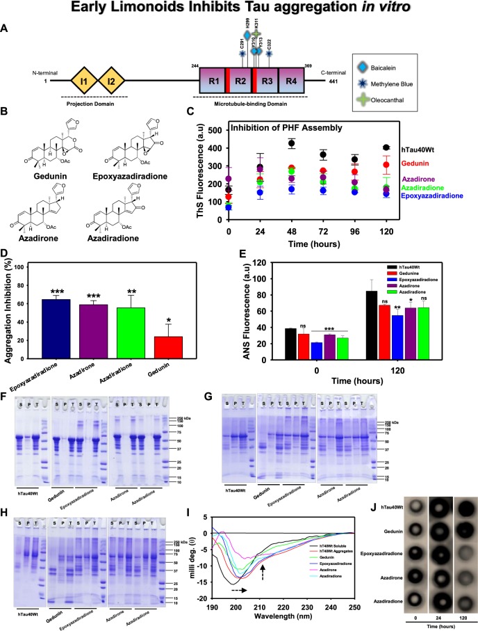
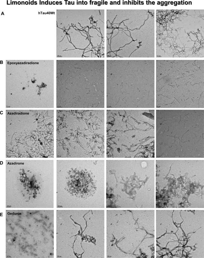
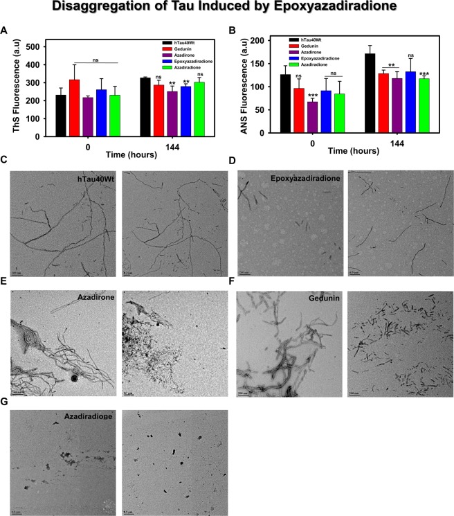
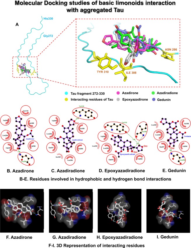
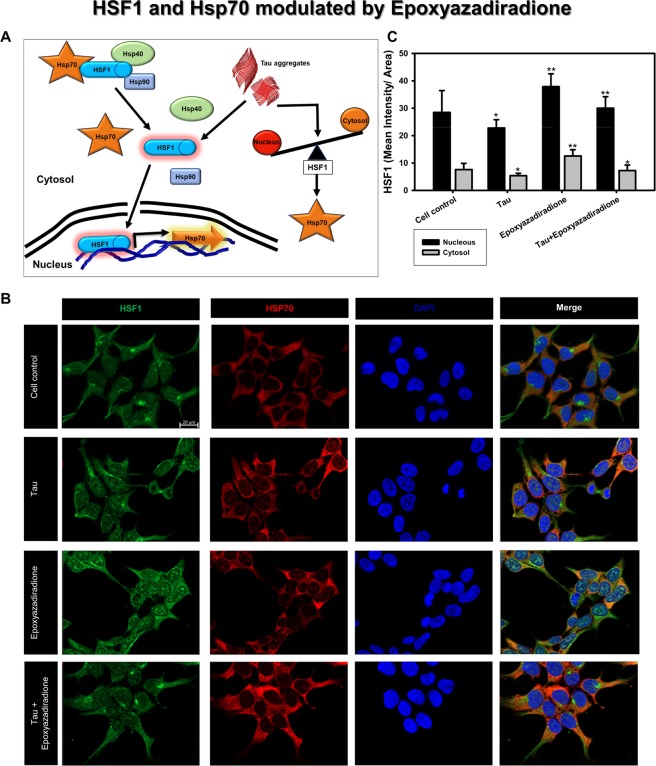
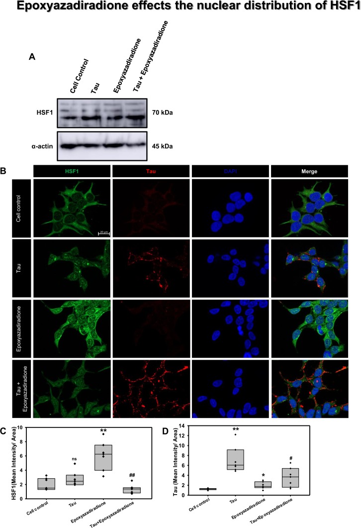
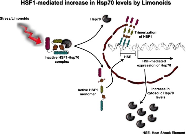
The Alzheimer's disease pathology is associated with accumulation of intracellular neurofibrillary tangles and extracellular senile plaques. The formation of initial nucleus triggers conformational changes in Tau and leads to its deposition. Hence, there is a need to eliminate these toxic proteins for proper functioning of neuronal cells. In this aspect, we screened the effect of basic limonoids such as gedunin, epoxyazadiradione, azadirone and azadiradione on inhibiting Tau aggregation as well as disintegration of induced Tau aggregates. It was observed that these basic limonoids effectively prevented aggregates formation by Tau and also exhibited the property of destabilizing matured Tau aggregates. The molecular docking analysis suggests that the basic limonoids interact with hexapeptide regions of aggregated Tau. Although these limonoids caused the conformational changes in Tau to β-sheet structure, the cytological studies indicate that basic limonoids rescued cell death. The dual role of limonoids in Tau aggregation inhibition and disintegration of matured aggregates suggests them to be potent molecules in overcoming Tau pathology. Further, their origin from a medicinally important plant neem, which known to possess remarkable biological activities was also found to play protective role in HEK293T cells. Basic limonoids were non-toxic to HEK293T cells and also aided in activation of HSF1 by inducing its accumulation in nucleus. Western blotting and immunofluorescence studies showed that HSF1 in downstream increased the transcription of Hsp70 thus, aggravating cytosolic Hsp70 levels that can channel clearance of aberrant Tau. All these results mark basic limonoids as potential therapeutic natural products.


Conflict of interest statement

The authors declare no competing interests.

Figures

Figure 1
Figure 1
The aggregation inhibition of Tau by basic limonoids. (A) Diagrammatic representation of full-length Tau comprising 441 amino acids with two inserts towards N-terminal and four repeats towards C-terminal. The inserts functions as projection domain, which serves to separate microtubule bundles. The four repeats act as microtubule-binding domain and helps in tubulin polymerization into microtubules. AD triggers pathological modifications in repeat region, leading to the loss of Tau affinity for microtubule. This causes destabilization of microtubule and aggregation of Tau. The bar diagram depicts the binding sites for various small molecules such as Baicalein, Methylene blue and Oleocanthal interacting with Tau. (B) Structure of the basic limonoids isolated from the fruit coat of Azadirachta indica. (C,D) Inhibition of full-length Tau aggregation by basic limonoids was analysed by ThS fluorescence assay which shows the potency of epoxyazadiradione over other limonoids, which was also represented as rate of inhibition in terms of percentage. (E) ANS fluorescence revealed the change of hydrophobicity content in Tau at time intervals of 0 and 120 hours, according to which epoxyazadiradione have persistently low hydrophobicity when compared to control and other limonoids. (F) The SDS-PAGE analysis showed the formation of higher order aggregates at 0 hour by epoxyazadiradione, azadirone and azadiradione, whereas full-length Tau, the control shows no signature of aggregation. The sedimentation assay revealed the presence of these aggregates in supernatant (S) and not in the pellet (P) which indicated that these were soluble aggregates. (G) After 24 hours of incubation the full-length Tau aggregated and this phenomenon was observed in control and test. This was witnessed by Tau present in the pellet (P) fraction obtained after sedimentation. Tau exhibited extensive degradation in presence of gedunin, whereas other limonoids showed Tau aggregation. (H) At 120 hours the Tau was completely aggregated and separated in the pellet (P). Gedunin exhibited mostly the degradation of soluble Tau although aggregation was also evidenced. Epoxyazadiradione, azadirone and azadiradione also exhibited Tau aggregation at varied extent. (I) The CD spectra of native Tau showed random coil conformation, whereas on aggregation the spectra signified β-sheet conformation. Similarly, in presence of limonoids the change in Tau conformation to β-sheet was observed. The basic limonoids exhibited difference in the spectrum intensity and shift. This could be due to the variation in the structure of limonoids which led Tau to attain varied conformations. (J) The aggregation of Tau was probed by K9JA reveals the initial aggregation by limonoids at 0 and 24 hours. Later at 120 hours of incubation shows the fully aggregated Tau in absence of limonoids. Epoxyazadiradione showed more potential in inhibiting aggregation, followed by Azadirone and Azadiradione. Gedunin had poor effects in preventing aggregates formation. The graph was plotted using SigmaPlot 10.0, from Systat Software, Inc., San Jose California USA, www.systatsoftware.com.
Figure 2
Figure 2
TEM analysis for Tau aggregation in presence of basic limonoids. (A) The negative staining of Tau aggregates are formed in vitro in presence of inducer exhibited polymorphic structure of Tau morphology. (B,C) Epoxyazadiradione and azadiradione inhibited Tau aggregation and limited the Tau aggregation to shorter length filaments. (D) Azadirone led to the formation of amorphous aggregates by Tau. (E) Tau aggregates exhibited change in morphology and twist in presence of gedunin. This result indicated that gedunin might have altered the mode of aggregation, but have no effect on rate of aggregation.
Figure 3
Figure 3
Disaggregation of pre-formed Tau aggregates by basic limonoids. (A,B) The role of basic limonoids in disaggregation of Tau was analysed by ThS and ANS fluorescence assay. The ThS and ANS fluorescence were evidenced to increase at 144 hours. This indicated that limonoids have no role in solubilizing the preformed Tau aggregates. (C) The aggregates of Tau exhibited the morphology of extended and thick filaments in the absence of limonoids. (D) Epoxyazadiradione destabilizes the pre-formed aggregates to shorter fragments. (E,F) Azadirone and gedunin altered the morphology of Tau aggregates and disintegrated them into shorter filaments. (G) Azadiradione completely destabilized Tau aggregates. The graph was plotted using SigmaPlot 10.0, from Systat Software, Inc., San Jose California USA, www.systatsoftware.com.
Figure 4
Figure 4
Molecular Docking studies of basic limonoids with aggregated Tau model (Gly272-His330). (A) shows the binding site for the ligands and the residues of Tau involved in hydrogen bond interactions. (B–E) represents the 2D-graph generated by LigPlot+ for hydrogen bond and hydrophobic interactions of the ligands with the Tau residues. (B) Azadirone, (C) Azadiradione, (D) Epoxyazadiradione, and (E) Gedunin. (F–I) represents the corresponding 3-dimensional view of the interaction visualized by PyMol. (F) Azadirone, (G) Azadiradione, (H) Epoxyazadiradione, and (I) Gedunin.
Figure 5
Figure 5
Cytotoxicity by basic limonoids on HEK293T cell line by MTT assay. (A) 104 cells/well were seeded in 96 well plate and incubated with 0.05 and 50 μM concentrations of basic limonoids at 37 °C, 5% CO2 for 24 hours, followed by MTT assay and calculation of percentage cell viability. The toxicity profile for four basic limonoids were: azadiradione>epoxyazadiradione>gedunin>azadirone. (B) HEK293T cells were seeded in 96 well plate and co-treated with 10 μM of full-length Tau aggregates along with six different concentrations (1, 2, 5, 10, 20, and 50 μM) of the limonoids and incubated for 24 hours at 37 °C, 5% CO2. MTT Assay determined cell viability where azadiradione and epoxyazadiradione at 1 μM concentration were more effective to reduce human Tau aggregates mediated toxicity as compared to azadirone and gedunin in HEK293T cells. (C) The toxicity of resulted Tau species in presence and absence of basic limonoids were analysed by cell viability assay. The viability of Tau treated cells decreased to 62%, whereas in presence of epoxyazadiradione less toxic species were formed resulting in 73% viability. Gedunin, azadirone and azadiradione formed toxic Tau species due to which the cell viability was reduced to 50%, 43% and 44% respectively. The graph was plotted using SigmaPlot 10.0, from Systat Software, Inc., San Jose California USA, www.systatsoftware.com.
Figure 6
Figure 6
HSF1 localization to nuclear compartment. (A) HSF1-Hsp70/40/90 complex are present as inactive form in the cytosol. During stress condition, HSF1 is signalled to dissociate from Hsps and allocated to nucleus for active transcription of Hsp70 in order to manage the cellular protein burden. The altered balance between nuclear vs. cytosolic HSF1 is the key determinant of controlling effective proteostasis in AD. (B) Tau aggregates induced the Hsp70 level in HEK293T cells while epoxyazadiradione has induced the HSF1 with reduced Hsp70 level, which indicates the feed-back control of cellular proteostasis signalling. (C) The quantification showed increase in nuclear as well as cytosolic levels of HSF1 in epoxyazadiradione treatment which is also maintained in Tau stressed cells. Quantification was carried out by Zen 2.3 software (https://www.zeiss.com/microscopy/int/products/light-microscopes/axio-observer-for-biology.html).
Figure 7
Figure 7
Activation of HSF1 by epoxyazadiradione. (A) Treatment of HEK293T cells with Tau and epoxyazadiradione exhibited no changes of HSF1 in western blotting analysis and its quantification. (B) Extracellular Tau aggregates reduced the cellular HSF1 level and epoxyazadiradione increased HSF1 level in cytosol and nucleus. Tau aggregates were found to adhere in the cell periphery whereas the intracellular Tau level was negligible in HEK293T cells. (C,D) IF quantification showed a significant increase in intracellular HSF1 and Tau aggregates were observed on the cellular periphery. Quantification was carried out by Zen 2.3 software (https://www.zeiss.com/microscopy/int/products/light-microscopes/axio-observer-for-biology.html).
Figure 8
Figure 8
Hsp70 levels aggravated on epoxyazadiradione treatment. (A) Hsp70, an indispensable chaperone involved in proteotoxic stress, its levels are increased when cells are exposed to Tau. (B) Tau aggregates increased the Hsp70 level in HEK293T cells as compared to cell control where the cells showed an extended morphology. (C) Epoxyazadiradione alone and together with Tau aggregates have increased the cellular Hsp70 levels, which indicates the induction of cellular proteostasis machinery in the context of extracellular protein burden. Quantification was carried out by Zen 2.3 software (https://www.zeiss.com/microscopy/int/products/light-microscopes/axio-observer-for-biology.html).
Figure 9
Figure 9
Proposed mechanism for HSF1-mediated clearance of Tau. In normal conditions HSF1 is present as an inactive monomer in cytosol. HSF1 in inactive state forms complex with Hsp70. Triggering of Tau-induced stress or treatment of cells with limonoids leads to dissociation of Hsp70 from HSF1. HSF1 monomer is now subjected to phosphorylation and is transported into nucleus. In nucleus, HSF1 trimerizes and interacts with heat shock element (HSE) present in the upstream of Hsp70 promoter. This leads to increase in expression of Hsp70 and thus enhancing the cytosolic levels of Hsp70.

Similar articles

Cited by

References

    1. Moya K, Benowitz L, Schneider G, Allinquant B. The amyloid precursor protein is developmentally regulated and correlated with synaptogenesis. Developmental Biology. 1994;161:597–603. doi: 10.1006/dbio.1994.1055. - DOI - PubMed
    1. Zou C, et al. Amyloid precursor protein maintains constitutive and adaptive plasticity of dendritic spines in adult brain by regulating D‐serine homeostasis. The EMBO Journal. 2016;35:2213–2222. doi: 10.15252/embj.201694085. - DOI - PMC - PubMed
    1. Hiltunen M, van Groen T, Jolkkonen J. Functional roles of amyloid-β protein precursor and amyloid-β peptides: evidence from experimental studies. Journal of Alzheimer’s Disease. 2009;18:401–412. doi: 10.3233/JAD-2009-1154. - DOI - PubMed
    1. Binder LI, Frankfurter A, Rebhun LI. The distribution of tau in the mammalian central nervous system. The Journal of Cell Biology. 1985;101:1371–1378. doi: 10.1083/jcb.101.4.1371. - DOI - PMC - PubMed
    1. Avila J, Lucas JJ, Perez M, Hernandez F. Role of tau protein in both physiological and pathological conditions. Physiological Reviews. 2004;84:361–384. doi: 10.1152/physrev.00024.2003. - DOI - PubMed

Publication types

MeSH terms