. 2020 Mar 4;10(1):4033.
doi: 10.1038/s41598-020-60640-z.

Iron redox pathway revealed in ferritin via electron transfer analysis

Affiliations

Iron redox pathway revealed in ferritin via electron transfer analysis

Peng Chen et al. Sci Rep. .

Abstract

Ferritin protein is involved in biological tissues in the storage and management of iron - an essential micro-nutrient in the majority of living systems. While there are extensive studies on iron-loaded ferritin, its functionality in iron delivery is not completely clear. Here, for the first time, differential pulse voltammetry (DPV) has been successfully adapted to address the challenge of resolving a cascade of fast and co-occurring redox steps in enzymatic systems such as ferritin. Using DPV, comparative analysis of ferritins from two evolutionary-distant organisms has allowed us to propose a stepwise resolution for the complex mix of concurrent redox steps that is inherent to ferritins and to fine-tune the structure-function relationship of each redox step. Indeed, the cyclic conversion between Fe3+ and Fe2+ as well as the different oxidative steps of the various ferroxidase centers already known in ferritins were successfully discriminated, bringing new evidence that both the 3-fold and 4-fold channels can be functional in ferritin.


Conflict of interest statement

The authors declare no competing interests.

Figures

Figure 1
Figure 1
Ferritin structure and related iron binding sites, (a) The crystal structure of a ferritin cage, with the 24-subunit ensemble spanning ~12.5 nm. Each subunit carries a ferroxidase center buried inside a four-helix bundle. (b) Three ferritin sub-units indicating the four-helix bundles delimiting a 3-fold channel and displaying metal ions (purple) at the location of the ion channel and the ferroxidase site (indicated with circles). (c) The Fe2+/Fe3+ binding sites: A and B constitute the ferroxidase center, and site C is a ferrihydrite nucleation/gateway site. The dashed lines represent the cross section of the 24-meric cage (shell). (d) A schematic representation of the A, B, and C sites, within a ferritin shell. All crystal structure representations are made in PyMol based on 2CIH.pdb.
Figure 2
Figure 2
Differential pulse voltammetry (DPV) was used for the interrogation of the redox characteristics of the ferritin and incorporates (a) the application of a voltage pulse (of typical magnitude Vp of ~50 mV, and applied for ~100 ms, in our experiments), superposed on a steadily increasing base voltage (Vbase). The difference in the electrical current before and after the voltage pulse is a measure of the Faradic response of the ferritin and is manifested through an electrical current peak corresponding to the relevant redox (reduction/oxidation) reactions. (b) DPV scans of FeCl2 in aqueous solution. Black, pH ~7, and Red, pH ~14. The redox peaks, observed in the cathodic scan, correspond broadly to the Fe3+ → Fe2+ reduction processes, and assigned to the Fe (OH)3/Fe (OH)2. In the reverse voltage scan, anodic redox peaks arise from the Fe2+ → Fe3+ oxidation processes and are assigned to the Fe2+/Fe (OH)3 (c) A comparative DPV with cathodic (c) and anodic (a) scans of ChF (in a phosphate buffer and NaCl) – outer curves, and the Buffer (without ChF) – inner curves. The subtraction of the Buffer scans from the observed signals from the combined ChF + Buffer yield a clearer delineation of the peaks belonging to the ChF alone. (d) Same raw data as from panel (c) but after Buffer background signal subtraction. This last format is how voltammograms will be presented in the remaining of this work.
Figure 3
Figure 3
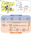
Redox kinetics in HuHF probed through differential pulse voltammetry and proposed redox mechanisms. (a) The HuHF ferroxidase center within a single subunit, indicating key residues and A, B and C sites. The respective distances between the metal ions are: A-B 3.5 Å, B-C 9.2 Å. PyMol was used to visualize HuHF with Zn in all 3 sites from 2CIH.pdb, despite the Glu27Asp mutation, (b) DPV derived peaks for HuHF, cathodic scan read first. The cathodic peak may be deconvolved to yield two contributions, related to the A and B sites of the ferritin, respectively, and the anodic peak is paired to the A peak. Roman numerals refer to relevant processes depicted in (c) - proposed mechanism for charge transfer processes in HuHF, as derived from the measured voltammograms. Orange arrows indicate the direction of electron flow, while green arrows indicate iron ion flow. The cathodic processes (reduction, top) at the two Fe3+ (in red) ions associated with the (II) A and (III) B sites yield two peaks corresponding to the creation of two Fe2+ ions (in blue) at the end of the cathodic scan. (IV) One of the formed Fe2+ is transported away from the ferritin and is (V) replaced by the Fe3+ from the C site or from the core (no specific proof of this was found in DPV). In the voltage-driven anodic process (bottom), (VI) the residual Fe2+ is oxidized to Fe3+ at site A only (when little Fe2+ is available) and constitutes the one peak observed in the anodic scan. (VII) The rearrangement of Fe3+ supplies the iron to the C site or to the core. Depending on the abundance of external free Fe2+ (limiting vs not limiting), the ferroxidase site can either remain vacant with limited free Fe2+ or take up iron and oxidize it when free Fe2+ is available abundantly, as shown in (VIII) and (IX). Three possible pathways have been indicated for the oxidation. The top path (in dashed lines) represents the fast, natural oxidation taking place inside the ferritin in the time frame between the cathodic and anodic scans. The two others are the voltage-driven reactions, associated with the voltammogram.
Figure 4
Figure 4
Redox kinetics in Chaetopterus ferritin (ChF) probed through differential pulse voltammetry and proposed iron redox mechanisms. (a) The ferroxidase center structure in ChF, showing all key residues including Ser141. The respective distances between the metal ions are: D-A 6.3 Å, A-B 3.4 Å, B-C 9.3 Å. ChF structure shown from 5WPN.pdb and visualized in PyMol. An additional metal binding site near the ferroxidase center is found in 5WPN and labeled as site D. (b) DPV derived peaks for ChF, in the cathodic and anodic scans. The cathodic peak is deconvolved to yield contributions related to the A and B sites of the ferritin. The anodic oxidation peak is associated with the B peak only. (c) The hypothesized mechanism for the charge transfer processes related to the Fe redox kinetics in ChF based on the DPV scan. The cathodic processes (top) at the two Fe3+ ions are associated successively with the (II) A and (III) B sites and yield two peaks corresponding to the Fe2+ and Fe3+ rearrangement at the end of the cathodic scan (IV), involving the D site. One of the formed Fe2+ ions is then transported away from the ferritin and (V) site A is occupied by Fe3+ from inside the ferritin. In the anodic oxidation process, (VI) the residual Fe2+ is oxidized to the Fe3+ in site B and constitutes the one peak observed in the anodic scans. (VII) The oxidized iron supplies the Fe3+ to the inner iron site. (VIII) and (IX) are follow-up steps when free Fe2+ is available.

Similar articles

Cited by

References

    1. Marchetti A, et al. Ferritin is used for the iron storage in bloom-forming marine pennate diatoms. Nature. 2009;457:467. doi: 10.1038/nature07539. - DOI - PubMed
    1. Liu X, Theil EC. Ferritins: Dynamic Management of Biological Iron and Oxygen Chemistry. Acc. Chem. Res. 2005;38:167–175. doi: 10.1021/ar0302336. - DOI - PubMed
    1. Arosio P, Levi S. Ferritin, iron homeostasis, and oxidative damage. Free Radic. Biol. Med. 2002;33:457–463. doi: 10.1016/S0891-5849(02)00842-0. - DOI - PubMed
    1. Smith JL. The physiological role of ferritin-like compounds in bacteria. Crit. Rev. Microbiol. 2004;30:173–185. doi: 10.1080/10408410490435151. - DOI - PubMed
    1. Camacho A, Waletr XA, Picazo A, Zopfi J. Photoferrotrophy: Remains of an Ancient Photosynthesis in Modern Environments. Front. Microbiol. 2017;8:323. doi: 10.3389/fmicb.2017.00323. - DOI - PMC - PubMed

Publication types