

Restenosis of a Polytetrafluoroethylene-Covered Stent Visualized by Coronary Angioscopy and Optical Coherence Tomography: A Case Report
- PMID: 32132819
- PMCID: PMC7054065
- DOI: 10.1055/s-0039-1685510
Restenosis of a Polytetrafluoroethylene-Covered Stent Visualized by Coronary Angioscopy and Optical Coherence Tomography: A Case Report
Abstract
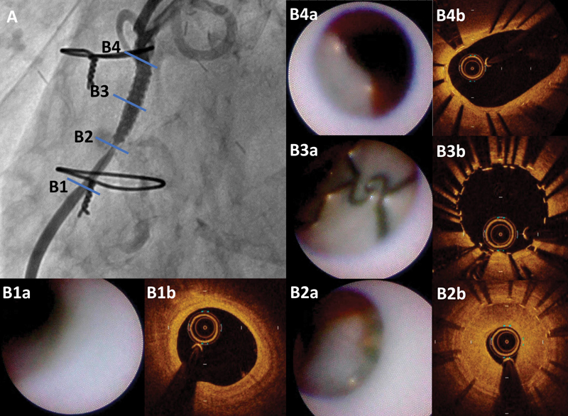
An expandable polytetrafluoroethylene (PTFE)-covered stent graft is beneficial for the treatment of coronary perforations. However, several reports have shown that restenosis and thrombotic occlusion occasionally occur in the stented segment after PTFE-covered stent implantation. A restenosis case after treatment with PTFE-covered stent against saphenous vein graft (SVG) perforation has never been evaluated with optical coherence tomography (OCT) or coronary angioscopy (CAS). This case report presents a 75-year-old man treated with a PTFE-covered stent after he suffered from SVG perforation 6 months ago. He was found to have a focal restenosis of the distal edge of the PTFE-covered stent and underwent percutaneous coronary intervention. OCT showed focal restenosis with homogeneous neointima and exposed struts in the middle and proximal part of the PTFE-covered stent. CAS showed white neointima with a smooth surface at the restenosis site and a sharp border against proximal exposed struts with characteristic links. This case study showed, for the first time in vivo and in a human, the neointimal characteristics of restenosis and uncovered stent struts in a PTFE-covered stent which had been implanted 6 months before. The delayed endothelialization was sustained until 12 months after implantation.
Keywords: OCT; PCI; case study; coronary angioscopy; optical coherence tomography; percutaneous coronary intervention; polytetrafluoroethylene-covered stent.
© Thieme Medical Publishers.
Conflict of interest statement
Conflict of Interest The authors declare that there is no conflict of interest.
Figures


References
-
- Ajluni S C, Glazier S, Blankenship L, O'Neill W W, Safian R D. Perforations after percutaneous coronary interventions: clinical, angiographic, and therapeutic observations. Cathet Cardiovasc Diagn. 1994;32(03):206–212. - PubMed
-
- Gruberg L, Pinnow E, Flood Ret al. Incidence, management, and outcome of coronary artery perforation during percutaneous coronary intervention Am J Cardiol 20008606680–682., A8 - PubMed
-
- Javaid A, Buch A N, Satler L F et al. Management and outcomes of coronary artery perforation during percutaneous coronary intervention. Am J Cardiol. 2006;98(07):911–914. - PubMed
-
- Shimony A, Zahger D, Van Straten M et al. Incidence, risk factors, management and outcomes of coronary artery perforation during percutaneous coronary intervention. Am J Cardiol. 2009;104(12):1674–1677. - PubMed
-
- Campbell P G, Hall J A, Harcombe A A, de Belder M A. The Jomed Covered Stent Graft for coronary artery aneurysms and acute perforation: a successful device which needs careful deployment and may not reduce restenosis. J Invasive Cardiol. 2000;12(05):272–276. - PubMed
Publication types
LinkOut - more resources
Full Text Sources
Miscellaneous
