. 2020 Feb 18;11(7):699-726.
doi: 10.18632/oncotarget.27487.

DNA dependent protein kinase (DNA-PK) enhances HIV transcription by promoting RNA polymerase II activity and recruitment of transcription machinery at HIV LTR

Affiliations

DNA dependent protein kinase (DNA-PK) enhances HIV transcription by promoting RNA polymerase II activity and recruitment of transcription machinery at HIV LTR

Sonia Zicari et al. Oncotarget. .

Abstract

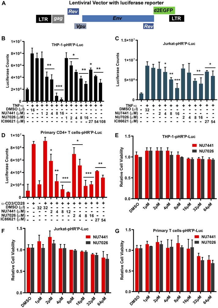
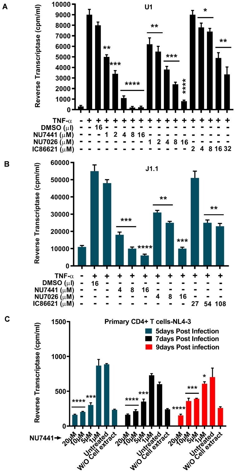
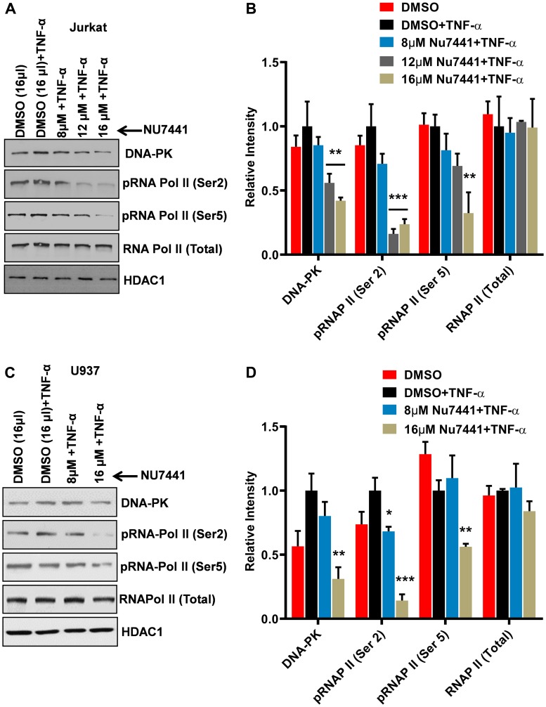
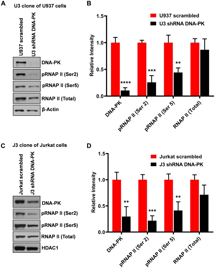
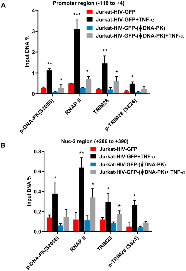
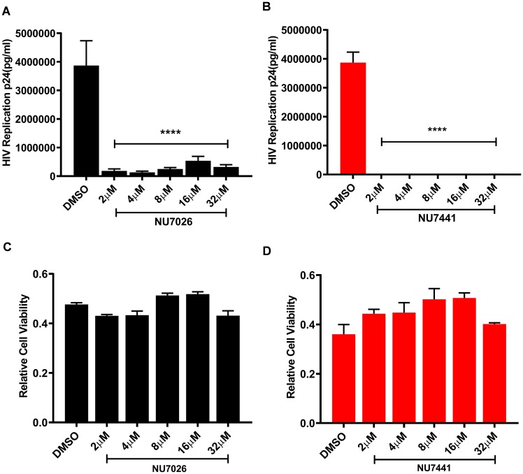
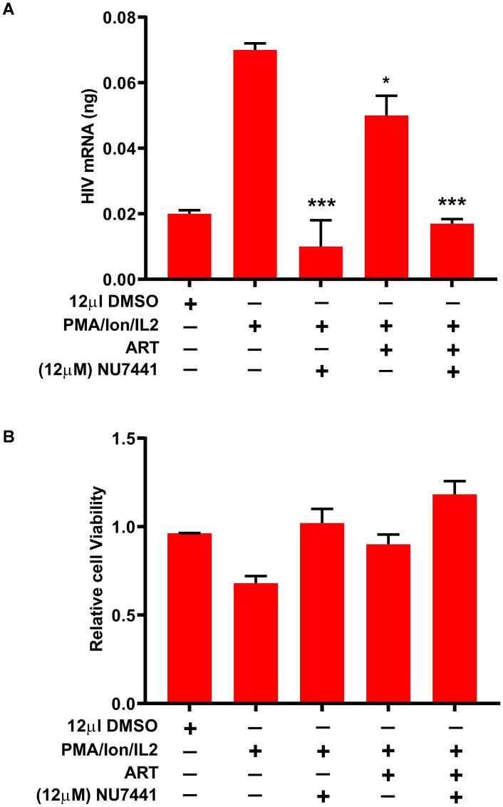
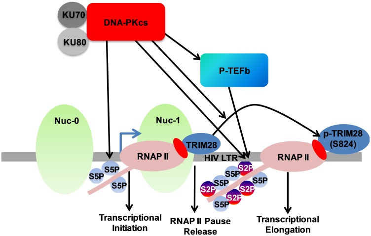
Despite reductions in mortality from the use of highly active antiretroviral therapy (HAART), the presence of latent or transcriptionally silent proviruses prevents HIV cure/eradication. We have previously reported that DNA-dependent protein kinase (DNA-PK) facilitates HIV transcription by interacting with the RNA polymerase II (RNAP II) complex recruited at HIV LTR. In this study, using different cell lines and peripheral blood mononuclear cells (PBMCs) of HIV-infected patients, we found that DNA-PK stimulates HIV transcription at several stages, including initiation, pause-release and elongation. We are reporting for the first time that DNA-PK increases phosphorylation of RNAP II C-terminal domain (CTD) at serine 5 (Ser5) and serine 2 (Ser2) by directly catalyzing phosphorylation and by augmenting the recruitment of the positive transcription elongation factor (P-TEFb) at HIV LTR. Our findings suggest that DNA-PK expedites the establishment of euchromatin structure at HIV LTR. DNA-PK inhibition/knockdown leads to the severe impairment of HIV replication and reactivation of latent HIV provirus. DNA-PK promotes the recruitment of Tripartite motif-containing 28 (TRIM28) at LTR and assists the release of paused RNAP II through TRIM28 phosphorylation. These results provide the mechanisms through which DNA-PK controls the HIV gene expression and, likely, can be extended to cellular gene expression, including during cell malignancy, where the role of DNA-PK has been well-established.

Keywords: DNA-PK; DNA-PK inhibitors; HIV; replication; transcription.


Conflict of interest statement

CONFLICTS OF INTEREST The authors declare that they have no conflicts of interests.

Figures

Figure 1
Figure 1. DNA-PK inhibitors repress HIV transcription without showing cytotoxicity.
(A) HIV- based lentiviral vector, pHR’P-Luc, which express luciferase reporter gene through HIV LTR promoter. (B) THP-1, a monocytic cell line, (C) Jurkat, a T cell line, and (D) primary CD4+ T cells carrying integrated latent pHR’P-Luc provirus in their genome were treated with indicated amounts of the DNA-PK inhibitors NU7441, NU7026, IC86621 or DMSO solvent-control along with or without 10 ng/ml of TNF-α. After 48 hours cells were lysed, and equal amount of proteins were used for luciferase assays. MTS cell viability assays were also performed after 5 days of culture (E) THP-1-pHR’P-Luc, (F) Jurkat-pHR’P-Luc and (G) primary CD4+ T cells-pHR’P-Luc. The results represent the Mean ± SD of three independent assays. The p value of statistical significance was set at either: p < 0.05 (*), 0.01 (**), 0.001 (***), or 0.0001 (****) when compared with DMSO treated stimulation as control.
Figure 2
Figure 2. DNA-PK inhibitors drastically impair latent proviral reactivation and HIV replication.
For assessing the impact of DNA-PK inhibitors on the latent proviral reactivation and replication, the cells were pre-incubated with indicated amounts of DNA-PK inhibitors or DMSO control, overnight. Next day, cells were supplied with fresh media containing inhibitors and/or 10 ng/ml TNF-α, accordingly, and further incubated for 48 hours. The cell supernatants were collected and used for the Reverse Transcriptase (RT) assays performed in (A) U1 (latently infected U937 based monocytic cell line), (B) J1.1 cell line (latently infected Jurkat based T cell line) and (C) Freshly infected primary CD4+ T cells. The results represent the mean ± SD of three independent assays. The statistical significance was set at: p < 0.05 (*), 0.01 (**), 0.001 (***), or 0.0001 (****) versus DMSO treated stimulated cells, as control.
Figure 3
Figure 3. DNA-PK inhibitors repress HIV transcription by restricting RNAP II CTD phosphorylation.
(A) Jurkat cells and (C) U937 cells were treated with increasing concentrations of the DNA-PK inhibitor NU7441 and then activated with TNF-α (10 ng/ml) for 3 hours. The cells were harvested, and nuclear extracts were assessed through western blot. The phosphorylation state of carboxyl terminal domain (CTD) of RNA polymerase II (RNAP II) was assessed through western blot using antibodies that are specific for RNAP II and its phosphorylated forms (Ser2 and Ser5). HDAC1 was used as loading control. The presented data is one representative western blot analysis out of three. Densitometric analyses were performed on western blot bands using ImageJ software for both (B) Jurkat cells and (D) U937 cells. The results represent the Mean ± SD of three different independent assays. Statistical significance is set as p < 0.05 (*), 0.01 (**), or 0.001 (***) compared to stimulated, but without DNA-PK inhibitor.
Figure 4
Figure 4. Knockdown of DNA-PK restricts RNAP II CTD phosphorylation.
The endogenous DNA-PK was knocked down using lentiviral vectors expressing shRNA sequences specific for DNA-PK. The vector expressing non-targeting scrambled shRNA sequence was used as control. Western blot assays showing nuclear levels of DNA-PK, total RNAP II, RNAP II (Ser2) and RNAP II (Ser5) in (A) U937 cells and corresponding DNA-PK knockdown clone U3 and (C) Jurkat cells and DNA-PK knockdown clone J3. β-Actin and HDAC1 were used as loading controls. Densitometric analyses were performed on (B) U937 cells and (D) Jurkat cells western blot’s bands using imageJ software. Error bars represent the SD of three separate experiments. Statistical significance was set as p < 0.05 (*), 0.01 (**), 0.001 (***) or 0.0001 (****) versus scrambled shRNA control.
Figure 5
Figure 5. DNA-PK facilitates the recruitment of P-TEFb at HIV LTR.
(A) Schematic diagram of HIV based lentiviral vector, which carry either mutated Tat (H13L) or wild-type Tat and express GFP as reporter. In these experiments VSV-G pseudotyped HIV, harboring wild-type Tat gene (HIV-GFP), was used. The DNA-PK knockdown (J3) or control Jurkat cells with scrambled shRNA were infected with HIV-GFP and the provirus was allowed to become latent. The ChIP assays were performed before and after reactivating the latent provirus with TNF-α (10 ng/ml) for 30 minutes. ChIP assays were done using indicated antibodies and two transcriptionally relevant regions of LTR were amplified using specific primer sets: (B) promoter region of the LTR; and (C) Nuc-2 region of the LTR. HIV DNA levels were calculated as percentages of the reaction input after background subtraction. Error bars represent the mean ± SD of four independent experiments and three separate real-time qPCR measurements. Statistical significance, p < 0.05 (*), 0.01 (**), or 0.001 (***), indicates enrichment at LTR following latent proviral reactivation, in both wild and DNA-PK knockdown conditions.
Figure 6
Figure 6. DNA-PK plays a vital role in HIV gene expression.
Flow cytometric analyses of freshly infected DNA-PK knockdown or control cells with either pHR’-PNL-H13LTat-d2GFP or pHR’-PNL-wildTat-d2GFP. (A) DNA-PK knockdown Jurkat clone J3 and control Jurkat with scrambled shRNA. (B) DNA-PK knockdown U937 cell clones, U2 and U3, and control cells with scrambled shRNA. After 48 hours post-infection, GFP positive cells were analyzed using a flow cytometer. The uninfected Jurkat and U937 were used as background control. (C) Tat protein levels in DNA-PK knockdown U2 and U3 clones infected with pHR’-PNL-wildTat-d2GFP analyzed via western blot. (D and E), the relative expression levels of HIV mRNA in DNA-PK knockdown and scrambled control cells were evaluated via RT-qPCR. The qPCR results represent the mean ± SD of three independent assays. Statistical significance, p < 0.001 (***), marks restriction to HIV gene expression, following DNA-PK knock down.
Figure 7
Figure 7. DNA-PK facilitates both the recruitment of transcription factors and establishment of euchromatin structures at HIV LTR.
ChIP analysis in Jurkat-pHR’P-Luc before and after activating latent HIV provirus with TNF-α in the absence or presence of DNA-PK inhibitor NU7441. The cellular chromatin was immunoprecipitated using indicated antibodies. The recovered DNA was analyzed using primer sets amplifying (A) the promoter region of the LTR, and (B) nucleosome-1 (Nuc-1) region of the LTR. Data is represented as percentages of the input nuclear extract after background subtraction. Results represent the mean ± SD of two independent experiments and three separate real-time qPCR measurements. Statistical analysis signifies the recruitment kinetics following reactivation of the latent provirus in the presence of NU7441 at HIV LTR; p < 0.05 (*).
Figure 8
Figure 8. DNA-PK catalyzes the phosphorylation of TRIM28.
Western blots were performed to evaluate the expression levels of Trim28 and phospho-Trim28 [p-TRIM28 (S824)] in (A) Jurkat cells treated with increasing concentration of the DNA-PK inhibitor NU7441 with or without TNF-α, and in (C) DNA-PK knockdown clone J3 and scrambled shRNA control. Densitometric analyses of (B) Jurkat cells and (D) DNA-PK knockdown J3 clone were performed using ImageJ software. Data is expressed as mean ± SD; n = 3. Significant differences in protein expression were evaluated; p < 0.05 (*), 0.01 (**), or 0.001 (***) against the respective controls.
Figure 9
Figure 9. DNA-PK facilitates TRIM28 recruitment at HIV LTR.
Recruitment kinetics of TRIM28 and p-TRIM28-(S824) were assessed using ChIP assays in J3 and scrambled control, and also before and after reactivating latent HIV provirus. (A) Primer set amplifying the promoter region of the LTR and (B) primers amplifying the downstream Nuc-2region of the LTR. Data represent the percentages of the input after background subtraction. Results depicts the mean ± SD of two independent experiments and three separate real-time qPCR measurements. Statistical significance, p < 0.05 (*) or 0.01 (**), marks the reactivation of latent HIV provirus in both wild or reduced DNA-PK conditions.
Figure 10
Figure 10. DNA-PK inhibitors completely block HIV replication in MT-4 T cells without showing cytotoxicity.
MT-4 cells were treated overnight with increasing concentrations of the DNA-PK inhibitors (A) NU7026 or (B) NU7441; DMSO was used as solvent control. Next day, the cells were infected with a replication competent ×4 virus (HIVLAI.04). After 3 days of culture, cells supernatants were evaluated for p24 with Luminex (Luminexcorp). Significant inhibition of HIV replication in presence of DNA-PK inhibitors (increasing dose) were evaluated; p < 0.0001 (****). The cytotoxicity of the inhibitors in MT-4 cells was assessed with MTT assay. No cytotoxicity was detected in cells treated with (C) NU7026 or (D) NU7441, compared to the DMSO control. All the results represent the mean ± SD of four independent measurements.
Figure 11
Figure 11. DNA-PK inhibitors restrict the reactivation of latent provirus in PBMCs of HIV-infected patients.
PBMCs from HIV-infected patients were treated with either ART alone, NU7441 alone or both together overnight. The day after, medium was replaced with fresh medium containing the drugs and the cells were stimulated for 3 days with PMA/ionomycin/IL-2 to reactivate latent HIV provirus present in PBMCs. (A) Cellular RNAs were isolated and evaluated via RT-qPCR. (B) Cell viability was examined by MTS assays. The results were reproduced 3 more times, by mixing PBMCs of 4 different patients each time. Presented data are the mean ± SD for four experiments. Statistical significance; p < 0.05 (*) or 0.001 (***) marks the restriction to reactivation of latent provirus in the presence of DNA-PK inhibitor.
Figure 12
Figure 12. DNA-PK facilitates HIV transcription by targeting multiple mechanisms.
In our model presented here we suggest that DNA-PK supports the initiation phase of transcription by catalyzing the phosphorylation of RNAP II CTD at Ser5. DNA-PK facilitates transcriptional elongation by enhancing phosphorylation of RNAP II CTD at Ser2, both via directly catalyzing and through the recruitment of P-TEFb at HIV LTR. DNA-PK relieves the RNAP II pausing by catalyzing the phosphorylation of TRIM28.

Similar articles

Cited by

References

    1. Chun TW, Davey RT Jr, Engel D, Lane HC, Fauci AS. Re-emergence of HIV after stopping therapy. Nature. 1999; 401:874–875. 10.1038/44755. - DOI - PubMed
    1. Davey RT Jr, Bhat N, Yoder C, Chun TW, Metcalf JA, Dewar R, Natarajan V, Lempicki RA, Adelsberger JW, Miller KD, Kovacs JA, Polis MA, Walker RE, et al.. HIV-1 and T cell dynamics after interruption of highly active antiretroviral therapy (HAART) in patients with a history of sustained viral suppression. Proc Natl Acad Sci U S A. 1999; 96:15109–15114. 10.1073/pnas.96.26.15109. - DOI - PMC - PubMed
    1. Karn J. The molecular biology of HIV latency: breaking and restoring the Tat-dependent transcriptional circuit. Curr Opin HIV AIDS. 2011; 6:4–11. 10.1097/COH.0b013e328340ffbb. - DOI - PMC - PubMed
    1. Mbonye U, Karn J. Control of HIV latency by epigenetic and non-epigenetic mechanisms. Curr HIV Res. 2011; 9:554–567. 10.2174/157016211798998736. - DOI - PMC - PubMed
    1. Siliciano RF, Greene WC. HIV latency. Cold Spring Harb Perspect Med. 2011; 1:a007096. 10.1101/cshperspect.a007096. - DOI - PMC - PubMed