. 2020 Feb 18:8:10.
doi: 10.1038/s41413-020-0087-2. eCollection 2020.

Melatonin alleviates intervertebral disc degeneration by disrupting the IL-1β/NF-κB-NLRP3 inflammasome positive feedback loop

Affiliations

Melatonin alleviates intervertebral disc degeneration by disrupting the IL-1β/NF-κB-NLRP3 inflammasome positive feedback loop

Fan Chen et al. Bone Res. .

Abstract

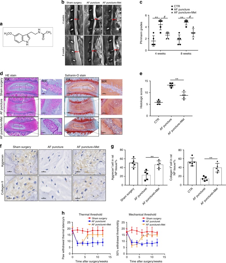
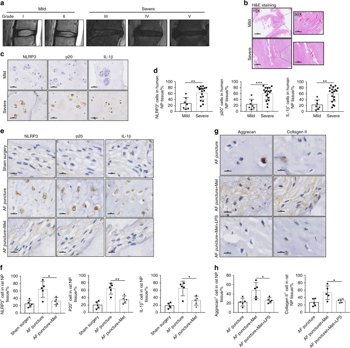
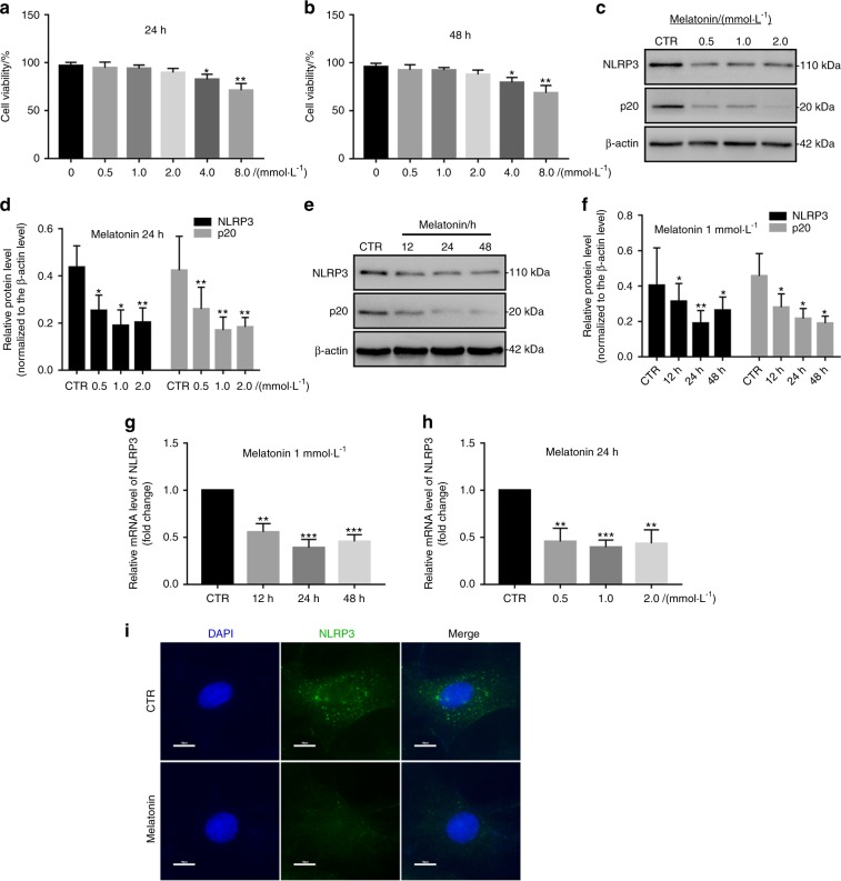
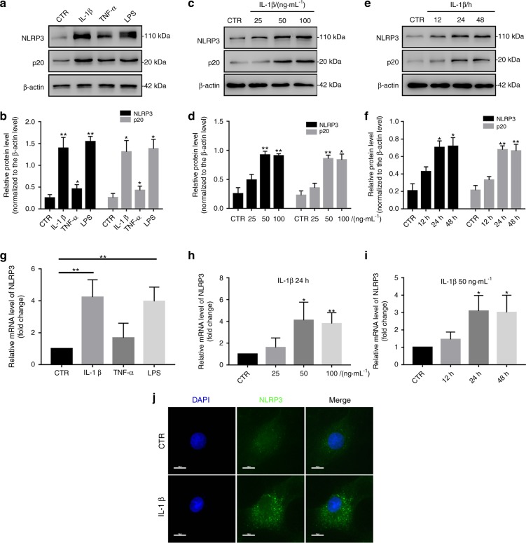
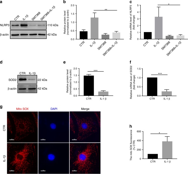
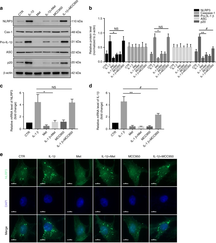
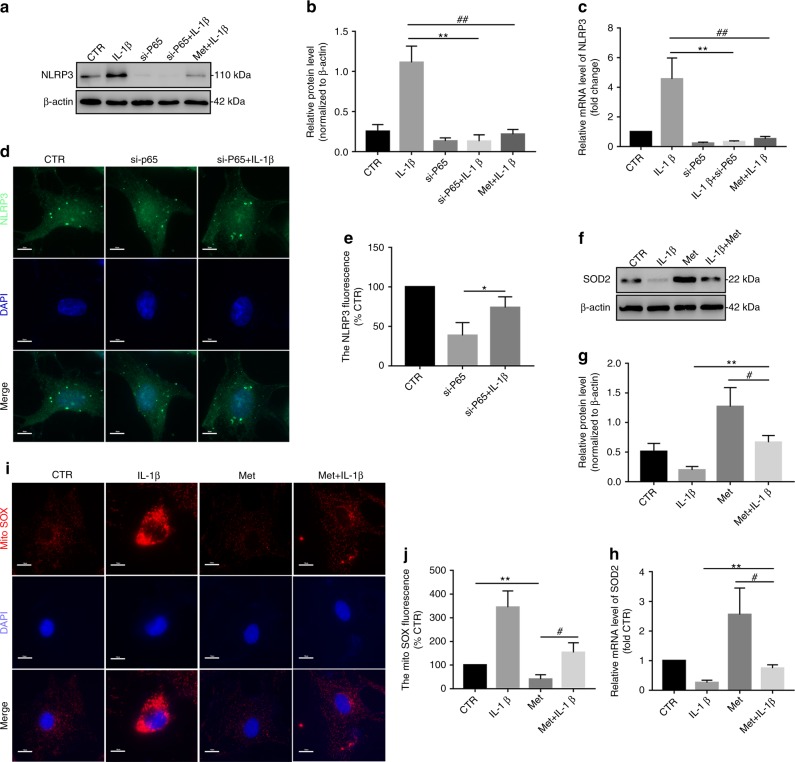
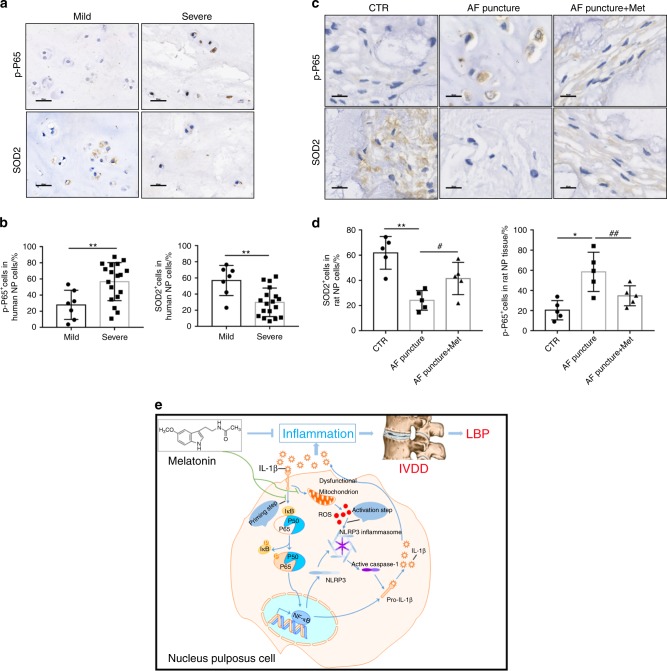
The inflammatory response is induced by the overexpression of inflammatory cytokines, mainly interleukin (IL)-1β, and is one of the main causes of intervertebral disc degeneration (IVDD). NLR pyrin domain containing 3 (NLRP3) inflammasome activation is an important source of IL-1β. As an anti-inflammatory neuroendocrine hormone, melatonin plays various roles in different pathophysiological conditions. However, its roles in IVDD are still not well understood and require more examination. First, we demonstrated that melatonin delayed the progression of IVDD and relieved IVDD-related low back pain in a rat needle puncture IVDD model; moreover, NLRP3 inflammasome activation (NLRP3, p20, and IL-1β levels) was significantly upregulated in severely degenerated human discs and a rat IVDD model. Subsequently, an IL-1β/NF-κB-NLRP3 inflammasome activation positive feedback loop was found in nucleus pulposus (NP) cells that were treated with IL-1β. In these cells, expression of NLRP3 and p20 was significantly increased, NF-κB signaling was involved in this regulation, and mitochondrial reactive oxygen species (mtROS) production increased. Furthermore, we found that melatonin disrupted the IL-1β/NF-κB-NLRP3 inflammasome activation positive feedback loop in vitro and in vivo. Melatonin treatment decreased NLRP3, p20, and IL-1β levels by inhibiting NF-κB signaling and downregulating mtROS production. Finally, we showed that melatonin mediated the disruption of the positive feedback loop of IL-1β in vivo. In this study, we showed for the first time that IL-1β promotes its own expression by upregulating NLRP3 inflammasome activation. Furthermore, melatonin disrupts the IL-1β positive feedback loop and may be a potential therapeutic agent for IVDD.

Keywords: Metabolism; Neurophysiology.


Conflict of interest statement

Competing interestsThe authors declare no competing interests.

Figures

Fig. 1
Fig. 1
Melatonin delays the progression of IVDD in vivo. a, b MRI images and Pfirrmann grade analysis of the rat models at 4 or 8 weeks after the operation. c, d H&E staining, Safranin-O staining and histologic score analysis of the rat models at 8 weeks after the operation (original magnification ×10, ×50; scale bar = 500 µm, 200 µm). e, f IHC staining and quantitative analysis of Aggrecan and Collagen II in IVDD rats treated with melatonin (original magnification ×400; scale bar = 50 µm). g Behavioral study of the rat models at different time points after the operation. CTR, Control. n = 5, *P < 0.05; **P < 0.01; #P < 0.05. The data are shown as the means ± SD
Fig. 2
Fig. 2
Melatonin alleviates IVDD by inhibiting NLRP3 inflammasome priming and activation in vivo. a MRI images of human discs. b H&E staining of human discs (original magnification ×10, ×50; scale bar = 500 µm, 100 µm). c, d IHC staining and quantitative analysis of NLRP3, P20, and IL-1β in human discs (original magnification ×400; scale bar = 50 µm). e, f IHC staining and quantitative analysis of NLRP3, P20, and IL-1β in IVDD rats treated with melatonin (original magnification ×400, scale bar = 50 µm). g, h IHC staining and quantitative analysis of Aggrecan and Collagen II in IVDD rats treated with melatonin or melatonin plus LPS (original magnification ×400; scale bar = 50 µm). n = 5, *P < 0.05; **P < 0.01; ***P < 0.001. The data are shown as the means ± SD
Fig. 3
Fig. 3
Melatonin inhibits NLRP3 inflammasome priming and activation in NP cells. a The chemical structure of melatonin. b, c NP cell viability after exposure to 0–8 mmol·L−1 NP for 24 h or 48 h. dg Western blot and quantitative analysis of NLRP3 and P20 in NP cells treated with different doses of melatonin or for different lengths of time. h, i RT-qPCR analysis of NLRP3 and P20 in NP cells treated with different doses of melatonin or at different time points. j IF analysis of NLRP3 in NP cells treated with melatonin (original magnification ×1 000; scale bar = 10 µm). CTR, Control. *P < 0.05; **P < 0.01. The data are shown as the means ± SD
Fig. 4
Fig. 4
IL-1β enhances NLRP3 inflammasome priming and activation in vitro. a, b Western blot and quantitative analysis of NLRP3 and P20 in NP cells treated with IL-1β, TNF-α or LPS. cf Western blot and quantitative analysis of NLRP3 and P20 in NP cells treated with different doses of IL-1β or at different time points. gi RT-qPCR analysis of NLRP3 in NP cells treated with IL-1β, TNF-α or LPS, different doses of IL-1β or at different time points. j IF analysis of NLRP3 in NP cells treated with IL-1β (original magnification ×1 000; scale bar = 10 µm). CTR, Control. *P < 0.05; **P < 0.01. The data are shown as the means ± SD
Fig. 5
Fig. 5
The IL-1β/NF-κB-NLRP3 inflammasome positive feedback loop is established in vitro. a, b Western blot and quantitative analysis of NLRP3 in NP cells treated with IL-1β, SM7368, or IL-1β plus SM7368. c RT-qPCR analysis of NLRP3 in NP cells treated with IL-1β, SM7368, or IL-1β plus SM7368. df Western blot and RT-qPCR analysis of SOD2 in NP cells treated with IL-1β. g, h MitoSOX red staining and quantitative analysis of NP cells treated with IL-1β (original magnification ×1 000; scale bar = 10 µm). CTR, Control. *P < 0.05; **P < 0.01; ***P < 0.001. The data are shown as the means ± SD
Fig. 6
Fig. 6
Melatonin disrupts the IL-1β-NLRP3 inflammasome positive feedback loop in vitro. a, b Western blot and quantitative analysis of NLRP3, Caspase-1, pro-IL-1β, ASC, and P20 in NP cells treated with IL-1β, melatonin, IL-1β plus melatonin, MCC950, and MCC950 plus IL-1β. c, d RT-qPCR analysis of NLRP3 and pro-IL-1β in NP cells treated with IL-1β, melatonin, IL-1β plus melatonin, MCC950, and MCC950 plus IL-1β. e IF analysis of NLRP3 in NP cells with different treatments (original magnification ×1 000; scale bar = 10 µm). CTR, Control; Met, melatonin; Met + IL-1β, melatonin plus IL-1β; NS, no statistical significance. *P < 0.05; **P < 0.01; #P < 0.05. The data are shown as the means ± SD
Fig. 7
Fig. 7
Melatonin disrupts the IL-1β-NLRP3 inflammasome positive feedback loop by downregulating NF-κB signaling and mtROS production. ac Western blot and RT-qPCR analysis of NLRP3 in NP cells cultured with IL-1β, si-P65, si-P65, and melatonin plus IL-1β. d IF analysis of NLRP3 in NP cells exposed to si-P65 and si-P65 plus IL-1β (original magnification ×1 000; scale bar = 10 µm). eg Western blot and RT-qPCR analysis of SOD2 in NP cells treated with IL-1β, melatonin, and melatonin plus IL-1β. h, i MitoSOX red staining and quantitative analysis of NP cells cultured with melatonin and melatonin plus IL-1β (original magnification ×1 000; scale bar = 10 µm). CTR, Control; Met, melatonin; Met + IL-1β, melatonin plus IL-1β. *P < 0.05; **P < 0.01; #P < 0.05; ##P < 0.01. The data are shown as the means ± SD
Fig. 8
Fig. 8
Melatonin disrupts the IL-1β/NF-κB-NLRP3 inflammasome positive feedback loop in vivo. ad IHC staining and quantitative analysis of p-P65 and SOD2 in human discs and rats at 8 weeks after the operation (original magnification ×400; scale bar = 50 µm). e Proposed schematic representation of melatonin disruption of the IL-1β/NF-κB-NLRP3 inflammasome positive feedback loop to alleviate IVDD in vivo and in vitro. CTR, Control; AF puncture + Met, AF puncture plus melatonin. n = 5. *P < 0.05; **P < 0.01; #P < 0.05; ##P < 0.01. The data are shown as the means ± SD

Comment in

  • Targeting IL-1β expression in IVDD.
    McHugh J. McHugh J. Nat Rev Rheumatol. 2020 Apr;16(4):188. doi: 10.1038/s41584-020-0403-7. Nat Rev Rheumatol. 2020. PMID: 32144404 No abstract available.

References

    1. Vlaeyen, J. W. S. et al. Low back pain. Nat. Rev. Dis.Primers4, 52 (2018). - PubMed
    1. Hoy D, Brooks P, Blyth F, Buchbinder R. The epidemiology of low back pain. Best. Pract. Res. Clin. Rheumatol. 2010;24:769–781. doi: 10.1016/j.berh.2010.10.002. - DOI - PubMed
    1. Maher C, Underwood M, Buchbinder R. Non-specific low back pain. Lancet. 2017;389:736–747. doi: 10.1016/S0140-6736(16)30970-9. - DOI - PubMed
    1. Hoy D, et al. The global burden of low back pain: estimates from the Global Burden of Disease 2010 study. Ann. Rheum. Dis. 2014;73:968–974. doi: 10.1136/annrheumdis-2013-204428. - DOI - PubMed
    1. Yang G, et al. Rapid health transition in China, 1990–2010: findings from the Global Burden of Disease Study 2010. Lancet. 2013;381:1987–2015. doi: 10.1016/S0140-6736(13)61097-1. - DOI - PMC - PubMed