A Method for Producing Highly Pure Magnetosomes in Large Quantity for Medical Applications Using Magnetospirillum gryphiswaldense MSR-1 Magnetotactic Bacteria Amplified in Minimal Growth Media
- PMID: 32133346
- PMCID: PMC7041420
- DOI: 10.3389/fbioe.2020.00016
A Method for Producing Highly Pure Magnetosomes in Large Quantity for Medical Applications Using Magnetospirillum gryphiswaldense MSR-1 Magnetotactic Bacteria Amplified in Minimal Growth Media
Abstract
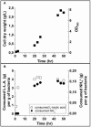
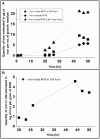
We report the synthesis in large quantity of highly pure magnetosomes for medical applications. For that, magnetosomes are produced by MSR-1 Magnetospirillum gryphiswaldense magnetotactic bacteria using minimal growth media devoid of uncharacterized and toxic products prohibited by pharmaceutical regulation, i.e., yeast extract, heavy metals different from iron, and carcinogenic, mutagenic and reprotoxic agents. This method follows two steps, during which bacteria are first pre-amplified without producing magnetosomes and are then fed with an iron source to synthesize magnetosomes, yielding, after 50 h of growth, an equivalent OD565 of ~8 and 10 mg of magnetosomes in iron per liter of growth media. Compared with magnetosomes produced in non-minimal growth media, those particles have lower concentrations in metals other than iron. Very significant reduction or disappearance in magnetosome composition of zinc, manganese, barium, and aluminum are observed. This new synthesis method paves the way towards the production of magnetosomes for medical applications.
Keywords: iron incorporation; magnetosome; magnetotactic bacteria; metals; minimal medium; yeast extract.
Copyright © 2020 Berny, Le Fèvre, Guyot, Blondeau, Guizonne, Rousseau, Bayan and Alphandéry.
Figures


Similar articles
-
Set-up of a pharmaceutical cell bank of Magnetospirillum gryphiswaldense MSR1 magnetotactic bacteria producing highly pure magnetosomes.Microb Cell Fact. 2024 Feb 28;23(1):70. doi: 10.1186/s12934-024-02313-4. Microb Cell Fact. 2024. PMID: 38419080 Free PMC article.
-
Effects of Environmental Conditions on High-Yield Magnetosome Production by Magnetospirillum gryphiswaldense MSR-1.Iran Biomed J. 2019 May;23(3):209-19. doi: 10.29252/.23.3.209. Epub 2019 Feb 24. Iran Biomed J. 2019. PMID: 30797225 Free PMC article.
-
Large-scale production of magnetosomes by chemostat culture of Magnetospirillum gryphiswaldense at high cell density.Microb Cell Fact. 2010 Dec 12;9:99. doi: 10.1186/1475-2859-9-99. Microb Cell Fact. 2010. PMID: 21144001 Free PMC article.
-
The bacterial magnetosome: a unique prokaryotic organelle.J Mol Microbiol Biotechnol. 2013;23(1-2):63-80. doi: 10.1159/000346543. Epub 2013 Apr 18. J Mol Microbiol Biotechnol. 2013. PMID: 23615196 Review.
-
Magnetosome biogenesis in magnetotactic bacteria.Nat Rev Microbiol. 2016 Sep 13;14(10):621-37. doi: 10.1038/nrmicro.2016.99. Nat Rev Microbiol. 2016. PMID: 27620945 Review.
Cited by
-
Imaging biomineralizing bacteria in their native-state with X-ray fluorescence microscopy.Chem Sci. 2025 Mar 3;16(26):12068-12079. doi: 10.1039/d4sc08375j. eCollection 2025 Jul 2. Chem Sci. 2025. PMID: 40474954 Free PMC article.
-
Parallel Manipulation and Flexible Assembly of Micro-Spiral via Optoelectronic Tweezers.Front Bioeng Biotechnol. 2022 Mar 21;10:868821. doi: 10.3389/fbioe.2022.868821. eCollection 2022. Front Bioeng Biotechnol. 2022. PMID: 35387303 Free PMC article.
-
Advances with metal oxide-based nanoparticles as MDR metastatic breast cancer therapeutics and diagnostics.RSC Adv. 2022 Nov 17;12(51):32956-32978. doi: 10.1039/d2ra02005j. eCollection 2022 Nov 15. RSC Adv. 2022. PMID: 36425155 Free PMC article. Review.
-
Bioinspired Magnetic Nanochains for Medicine.Pharmaceutics. 2021 Aug 16;13(8):1262. doi: 10.3390/pharmaceutics13081262. Pharmaceutics. 2021. PMID: 34452223 Free PMC article. Review.
-
Defining Local Chemical Conditions in Magnetosomes of Magnetotactic Bacteria.J Phys Chem B. 2022 Apr 14;126(14):2677-2687. doi: 10.1021/acs.jpcb.2c00752. Epub 2022 Apr 1. J Phys Chem B. 2022. PMID: 35362974 Free PMC article.
References
-
- Alphandéry E., Idbaih A., Adam C., Delattre J.-Y., Schmitt C., Guyot F., et al. . (2017). Development of non-pyrogenic magnetosome minerals coated with poly-l-lysine leading to full disappearance of intracranial U87-Luc glioblastoma in 100% of treated mice using magnetic hyperthermia. Biomaterials 141, 210–222. 10.1016/j.biomaterials.2017.06.026 - DOI - PubMed
-
- Amor M., Busigny V., Louvat P., Tharaud M., Gélabert A., Cartigny P., et al. (2018). Iron uptake and magnetite biomineralization in the magnetotactic bacterium Magnetospirillum magneticum strain AMB-1: an iron isotope study. Geochim Cosmochim. Acta 232, 225–243. 10.1016/j.gca.2018.04.020 - DOI
LinkOut - more resources
Full Text Sources
Other Literature Sources
Molecular Biology Databases

