. 2020 Feb 20:4:4.
doi: 10.1038/s41538-020-0064-6. eCollection 2020.

Annexin A1 accounts for an anti-inflammatory binding target of sesamin metabolites

Affiliations

Annexin A1 accounts for an anti-inflammatory binding target of sesamin metabolites

Yasuaki Kabe et al. NPJ Sci Food. .

Abstract

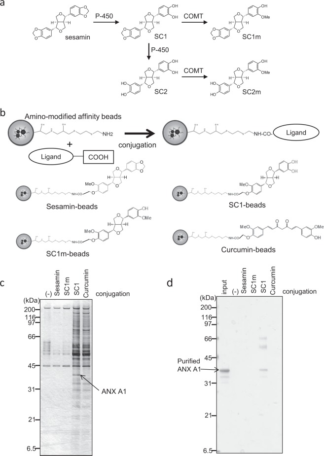
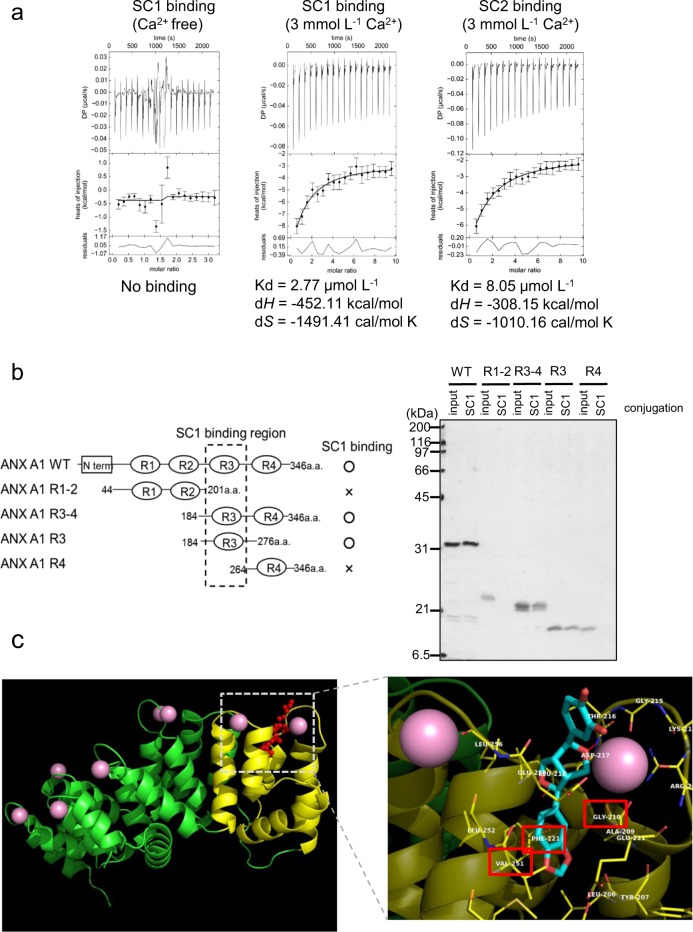
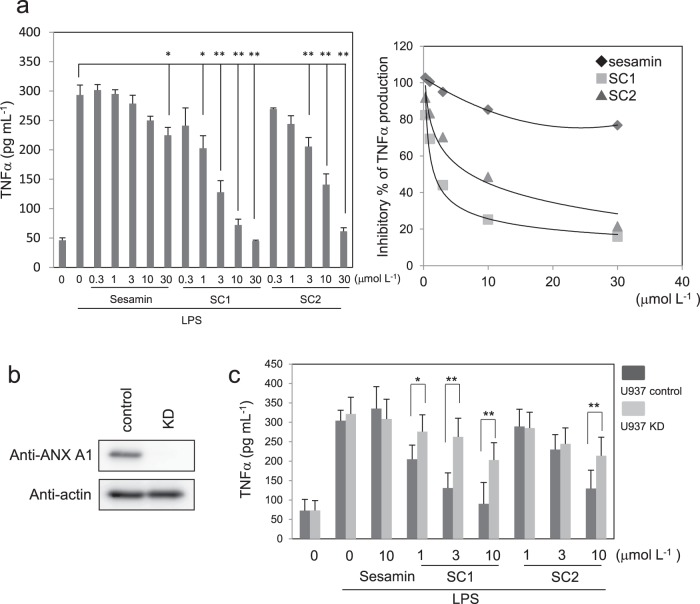
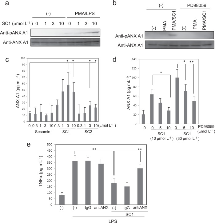
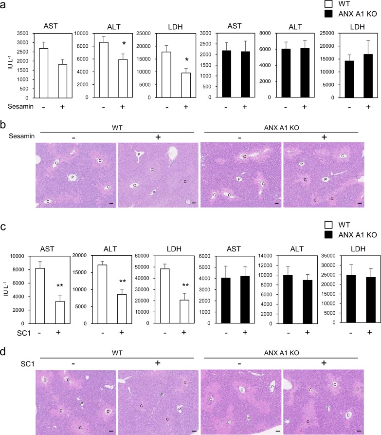
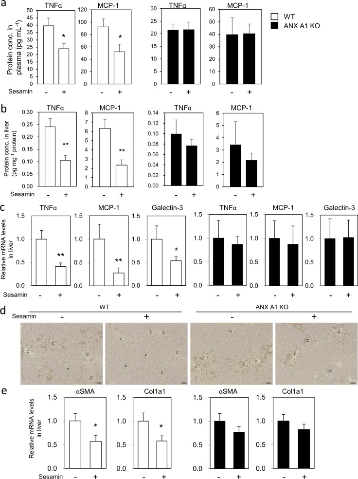
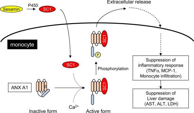
Sesamin [(7α,7'α,8α,8'α)-3,4:3',4'-bis(methylenedioxy)-7,9':7',9-diepoxylignane] is a major lignan in sesame seeds. Sesamin is converted to the catechol metabolite, SC1 [(7α,7'α,8α,8'α)-3',4'-methylenedioxy-7,9':7',9-diepoxylignane-3,4-diol] with anti-inflammatory effects after oral administration. However, its molecular target remains unknown. Analysis using high-performance affinity nanobeads led to the identification of annexin A1 (ANX A1) as an SC1-binding protein. SC1 was found to bind to the annexin repeat 3 region of ANX A1 with a high-affinity constant (Kd = 2.77 μmol L-1). In U937 cells, SC1 exhibited an anti-inflammatory effect dependent on ANX A1. Furthermore, administration of sesamin or SC1 attenuated carbon tetrachloride-induced liver damage in mice and concurrently suppressed inflammatory responses dependent on ANX A1. The mechanism involved SC1-induced ANX A1 phosphorylation at serine 27 that facilitates extracellular ANX A1 release. Consequently, the ANX A1 released into the extracellular space suppressed the production of tumor necrosis factor α. This study demonstrates that ANX A1 acts as a pivotal target of sesamin metabolites to attenuate inflammatory responses.

Keywords: Mechanism of action; Proteins.


Conflict of interest statement

Competing interestsD.T., Y.O., S.A., Y.K., T.R. and H.S. are employees of Suntory Wellness Ltd, which is a manufacturer of foods that contain sesamin. All authors declare no other competing interests.

Figures

Fig. 1
Fig. 1. Identification of ANX A1 as a sesamin metabolite-binding protein using affinity nanobeads.
a Scheme of sesamin metabolism in the liver. Sesamin is catalysed into SC1 and subsequently SC2 by cytochromes P450. SC1 or SC2 is further methylated into SC1m or SC2m by catechol O-methyl transferase (COMT). b Scheme of sesamin derivative-conjugation with affinity nanobeads. The carboxylated derivatives are conjugated with amino-modified nanobeads (upper panel). Lower panels show the chemical structures of the beads conjugating with sesamin, SC1, SC1m, or curcumin. c Affinity purification of the SC1-binding protein from U937 cell lysate. The affinity nanobeads conjugating with the ligands were incubated with U937 cell lysate, and bound proteins were analysed with SDS-PAGE before visualising using silver staining. The indicated protein (arrow) was identified as ANX A1 using peptide sequencing via ESI-MS. d Recombinant ANX A1 protein was incubated with affinity nanobeads, and bound proteins were analysed using SDS-PAGE before visualising with silver staining.
Fig. 2
Fig. 2. Analyses of binding property between ANX A1 and sesamin metabolites.
a Analyses of binding affinity between ANX A1 and sesamin metabolites using isothermal titration calorimetry (ITC). The binding affinity between ANX A1 and SC1 with ITC was analysed in the presence of EDTA (Ca2+ free, left panel) or CaCl2 (middle panel). Right panel showed the binding affinity between ANX A1 and SC2 using CaCl2-containing buffer. The binding values (Kd, ΔH, or ΔS) were calculated using the SEDPHAT program, and the data were shown as the average of three independent experiments. b Identification of the SC1-binding domain of ANX A1. The SC1-conjugating beads were incubated with wild-type (WT) or deletion mutant proteins of ANX A1 (left panel). Bound proteins were analysed with SDS-PAGE before visualising using silver staining (right panel). c Docking simulation of ANX A1 and SC1. ANX A1 is depicted as green or yellow (repeat 3 domain) ribbon (left panel). Ca2+ is depicted as pink balls. SC1 is shown as red sticks and balls. Close-up view of binding pocket of SC1 (light blue) at the Ca2+-binding region of ANX A1 repeat 3 domain (right panel). The amino acid residues marked by red squares (Gly210, Phe221 or Val251) indicate sites used for binding analysis of ANX A1 point mutants (Supplementary Fig. 3).
Fig. 3
Fig. 3. SC1 suppresses TNFα production dependent on ANX A1 in U937 cells.
a Suppressive effect of sesamin derivatives on TNFα production in U937 cells. U937 cells were differentiated by adding PMA for 24 h and treating with or without 1 µg mL−1 LPS and/or the indicated amount of sesamin, SC1 or SC2 for 12 h. TNFα production was analysed using ELISA. Right panel shows the relative inhibitory percentage of TNFα production by sesamin derivatives. b Protein expression of ANX A1 knockdown (KD) U937 cells. Control and shRNA for ANX A1 introducing lentivirus were infected into U937 cells. The cell lysates were analysed using western blotting using antibody against ANX A1 or β-actin. c U937 cells (control or ANX A1 KD) were differentiated with PMA and treated with or without 1 µg mL−1 LPS and/or the indicated amount of sesamin, SC1 or SC2. TNFα production was analysed using ELISA. All data represent the mean ± SD (n = 3). *p < 0.05, **p < 0.01 using unpaired Student’s t test.
Fig. 4
Fig. 4. SC1 elicits an anti-inflammatory effect by promoting the phosphorylation and extracellular release of ANX A1.
a, b U937 cells treated with or without PMA and LPS were incubated with the indicated amount of SC1 for 12 h (a), and the cell lysates were analysed using western blotting with antibody against ANX A1 or phosphorylated (Ser27) ANX A1. MAPK inhibitor PD98059 was treated 1 h prior to LPS treatment (b). c, d Extracellular release of ANX A1 from U937 cells was detected using the ELISA assay as mentioned above. PD98059 was treated 1 h prior to LPS treatment (d). e U937 cells were treated with control IgG or anti-ANX A1 antibody 1 h before LPS treatment in the presence or absence of 10 μmol L−1 SC1. The TNFα production in cell culture media was analysed using ELISA. All data represent the mean ± SD (n = 3). *p < 0.05, **p < 0.01 using unpaired Student’s t test.
Fig. 5
Fig. 5. ANX A1 is required for the hepato-protective effects of sesamin and SC1 in the CCl4-induced liver damage model.
a, b Effect of sesamin administration on the CCl4-induced liver damage in WT or ANX A1-KO mice. WT or ANX A1-KO mice (C57BL/6J) were intraperitoneally administered CCl4, and sesamin (25 mg kg−1 body weight), or vehicle was orally administered twice at 1 h before and 7 h after CCl4 administration. Hepatic damage marker enzymes (AST, ALT or LDH) in the plasma were measured 24 h after CCl4 administration (a). Data represent the mean ± SE. n= 13–15 mice in each group. *p < 0.05 using unpaired Student’s t test. The liver section was histologically evaluated using H&E staining (b). P and C indicated the portal and central vein, respectively. Scale bar: 50 μm. c, d Effect of SC1 on the CCl4-induced liver damage in WT or ANX A1-KO mice. SC1 (50 mg kg−1 body weight) or vehicle was intraperitoneally administered twice at 1 h before and 7 h after CCl4 administration. AST, ALT and LDH in the plasma, 24 h after CCl4 administration, were measured (c). Data represent the mean ± SE. n= 9–14 mice in each group. **p < 0.01 using unpaired Student’s t test. (d) The liver section was histologically evaluated using H&E staining. Scale bar: 50 μm.
Fig. 6
Fig. 6. ANX A1 is required for the suppressive effects of sesamin against CCl4-induced liver inflammation.
Effect of sesamin on CCl4–induced inflammation induced in WT or ANX A1-KO mice. WT or ANX A1-KO mice were intraperitoneally injected with CCl4. Sesamin (25 mg kg−1 body weight) or vehicle was administered twice at 1 h before and 7 h after CCl4 administration. The plasma or liver were collected for analysis 12 h after CCl4 administration. Production of TNFα and MCP-1 protein in the plasma (a) or in the liver (b) was analysed using ELISA. c Expression of TNFα, MCP-1, and galectin-3 mRNA in the liver was measured using qPCR, and normalised to 18S ribosomal RNA expression. The graph showed as the relative fold change compared to mRNA expression in the liver treated with vehicle. Data represent the mean ± SE. n = 5-10 mice in each group. *p < 0.05 or **p < 0.01 using unpaired Student’s t test. d The liver section collected 24 h after CCl4 administration with or without sesamin treatment was stained using galectin-3 antibody. Scale bar: 50 μm. e mRNA expression of αSMA or Col1a1 was measured by qPCR from the liver tissues collected 24 h after CCl4 administration with or without treatment of sesamin, and normalised by the expression of 18S rRNA. Data represent the mean ± SE. n = 13–15 mice in each group. *p < 0.05 using unpaired Student’s t test.
Fig. 7
Fig. 7. Schematic representation of the anti-inflammatory effect of SC1 mediated by ANX A1 activation.
SC1 is produced by metabolising sesamin in hepatocytes. In monocytes, SC1 directly binds to the repeat 3 domain of Ca2+ form ANX A1. Under stimulated conditions like PMA/LPS in monocytes, SC1 promotes the phosphorylation of ANX A1 Ser27 and the subsequent extracellular release of ANX A1, and thereby eliciting an anti-inflammatory effect via the suppression of TNFα or MCP-1 production.

Similar articles

Cited by

References

    1. Hamedi A, Zarshenas MM, Sohrabpour M, Zargaran A. Herbal medicinal oils in traditional Persian medicine. Pharm. Biol. 2013;51:1208–1218. doi: 10.3109/13880209.2013.777462. - DOI - PubMed
    1. Ma JQ, Ding J, Zhang L, Liu CM. Hepatoprotective properties of sesamin against CCl4 induced oxidative stress-mediated apoptosis in mice via JNK pathway. Food Chem. Toxicol. 2014;64:41–48. doi: 10.1016/j.fct.2013.11.017. - DOI - PubMed
    1. Chen X, Ying X, Chen L, Zhang W, Zhang Y. Protective effects of sesamin on liver fibrosis through antioxidative and anti-inflammatory activities in rats. Immunopharmacol. Immunotoxicol. 2015;37:465–472. doi: 10.3109/08923973.2015.1085064. - DOI - PubMed
    1. Yashaswini PS, Sadashivaiah B, Ramaprasad TR, Singh SA. In vivo modulation of LPS induced leukotrienes generation and oxidative stress by sesame lignans. J. Nutr. Biochem. 2017;41:151–157. doi: 10.1016/j.jnutbio.2016.12.010. - DOI - PubMed
    1. Cui Y, et al. Sesamin inhibits bacterial formylpeptide-induced inflammatory responses in a murine air-pouch model and in THP-1 human monocytes. J. Nutr. 2010;140:377–381. doi: 10.3945/jn.109.117804. - DOI - PubMed