. 2020 Jul;50(7):1067-1077.
doi: 10.1002/eji.201948257. Epub 2020 Apr 6.

Inhibition of stearoyl-CoA desaturases suppresses follicular help T- and germinal center B- cell responses

Affiliations

Inhibition of stearoyl-CoA desaturases suppresses follicular help T- and germinal center B- cell responses

Young Min Son et al. Eur J Immunol. 2020 Jul.

Abstract

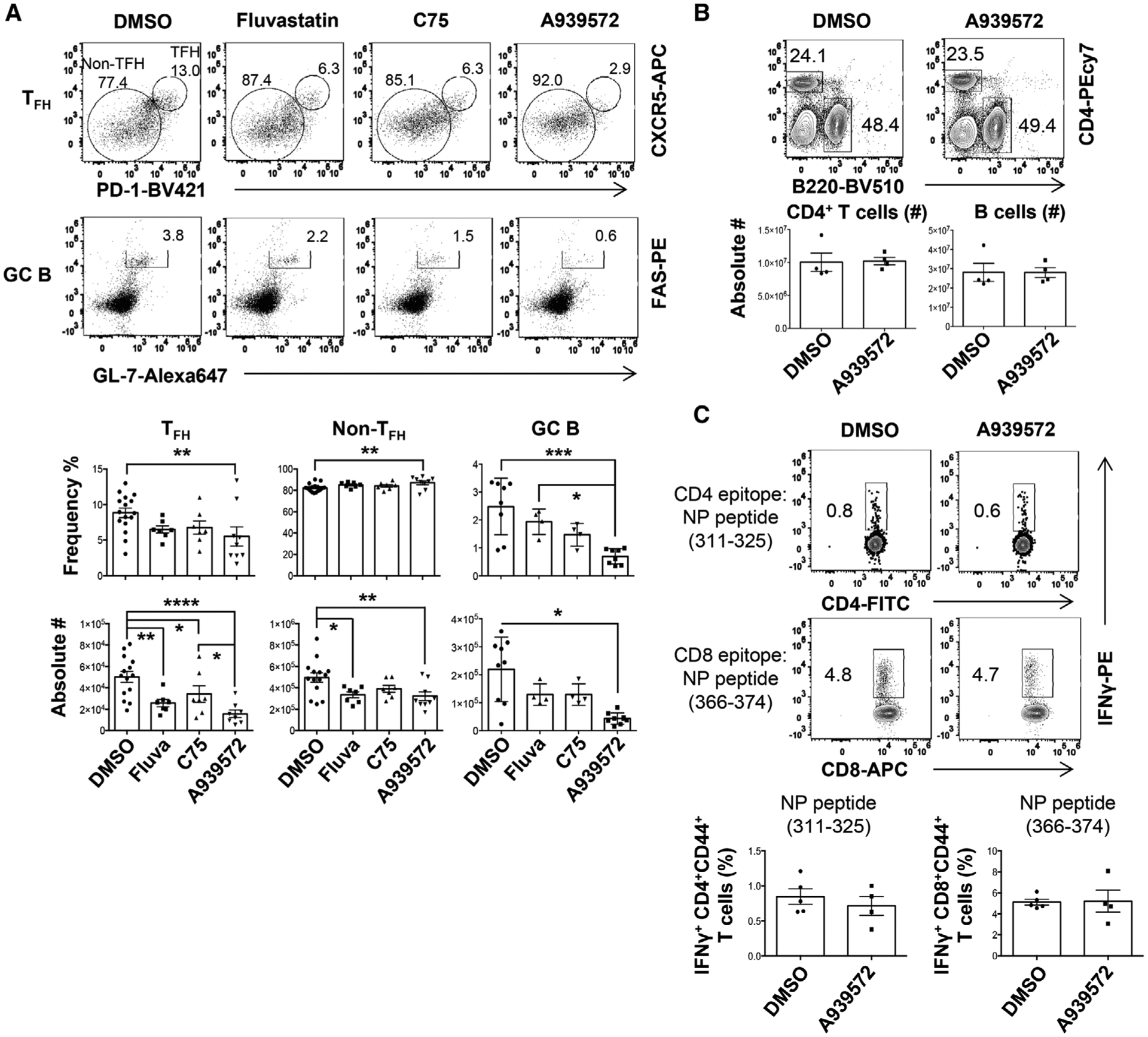
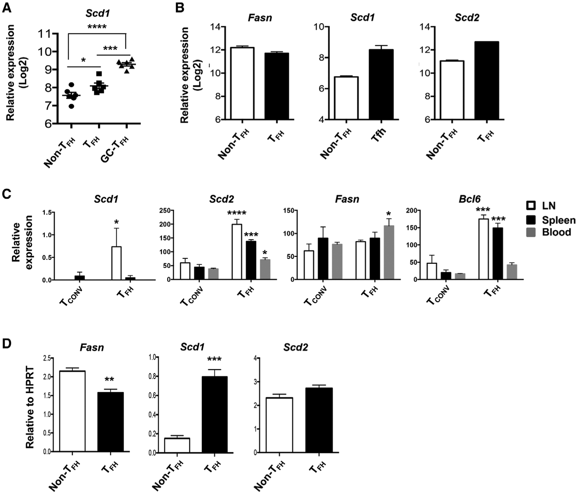
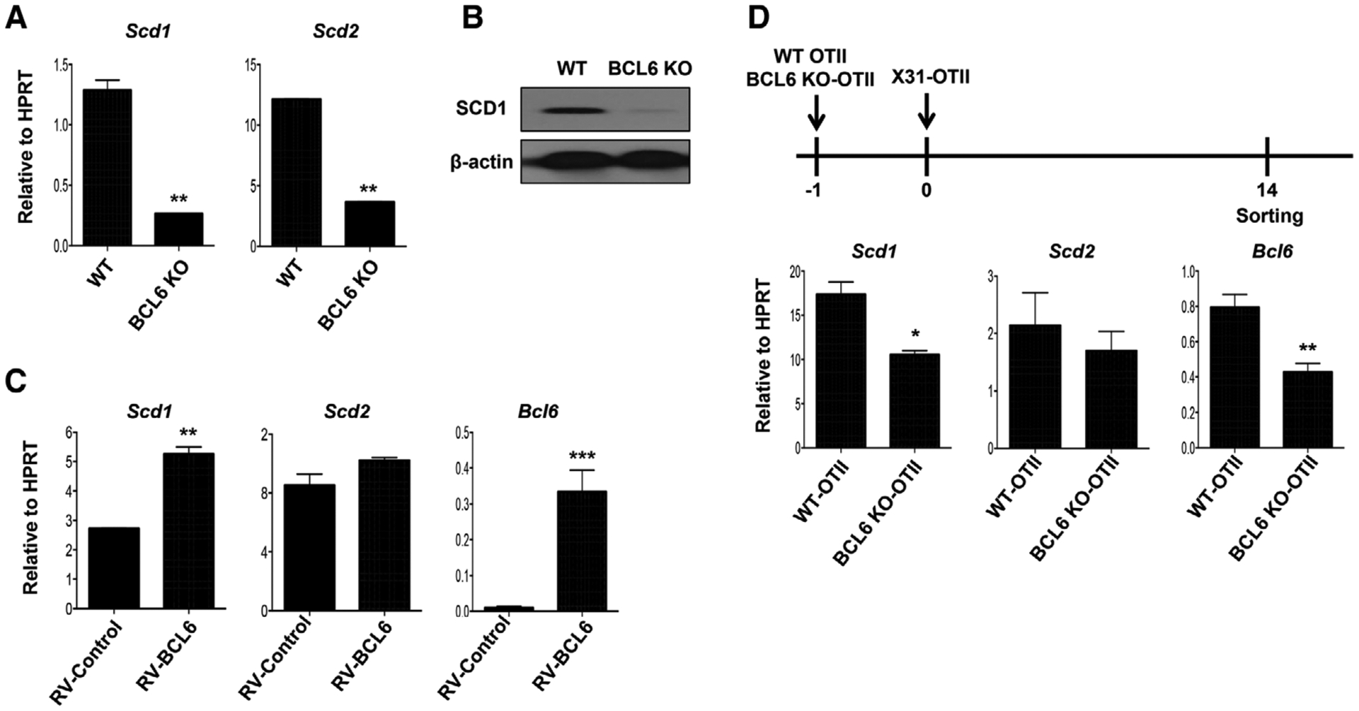
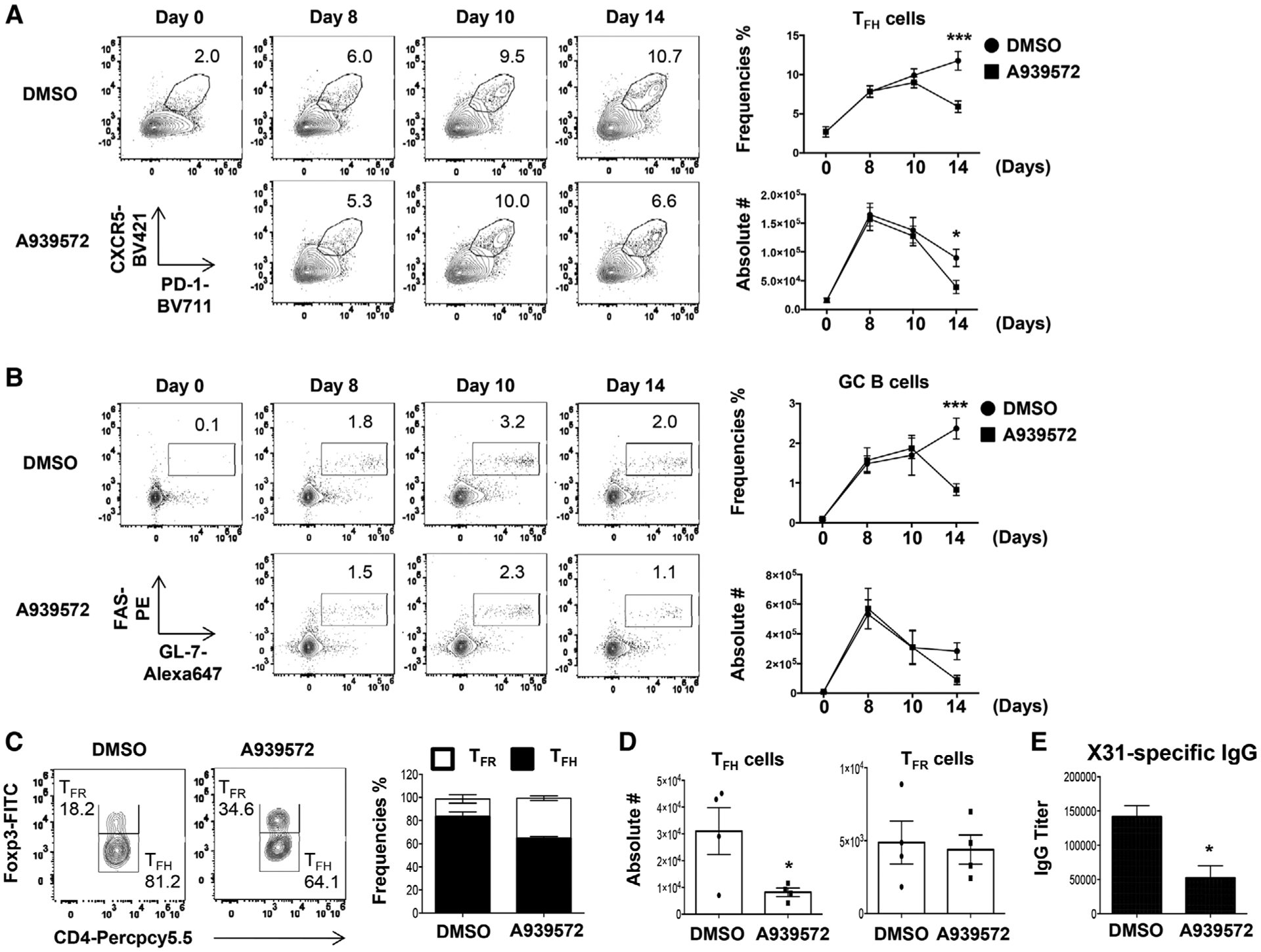
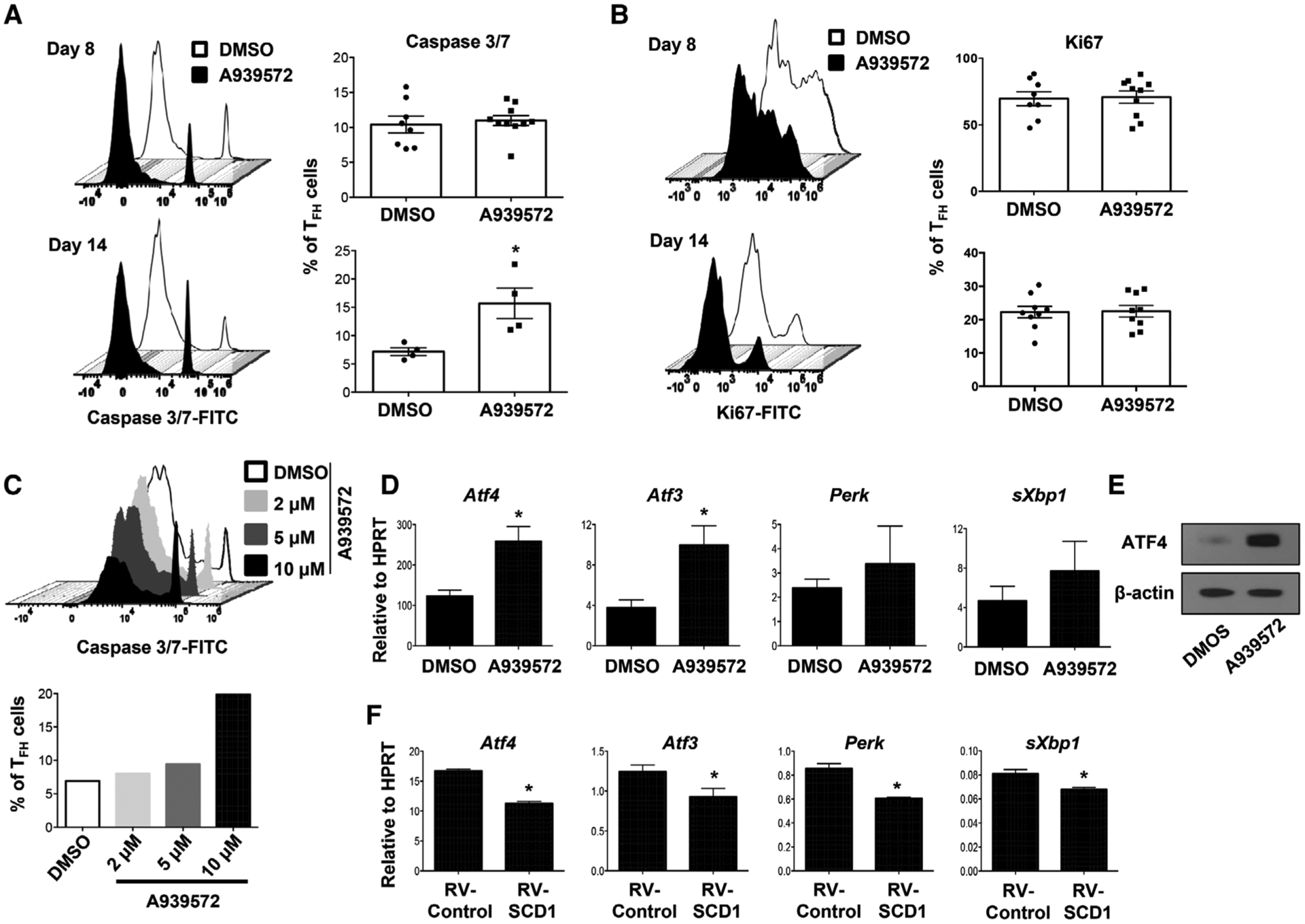
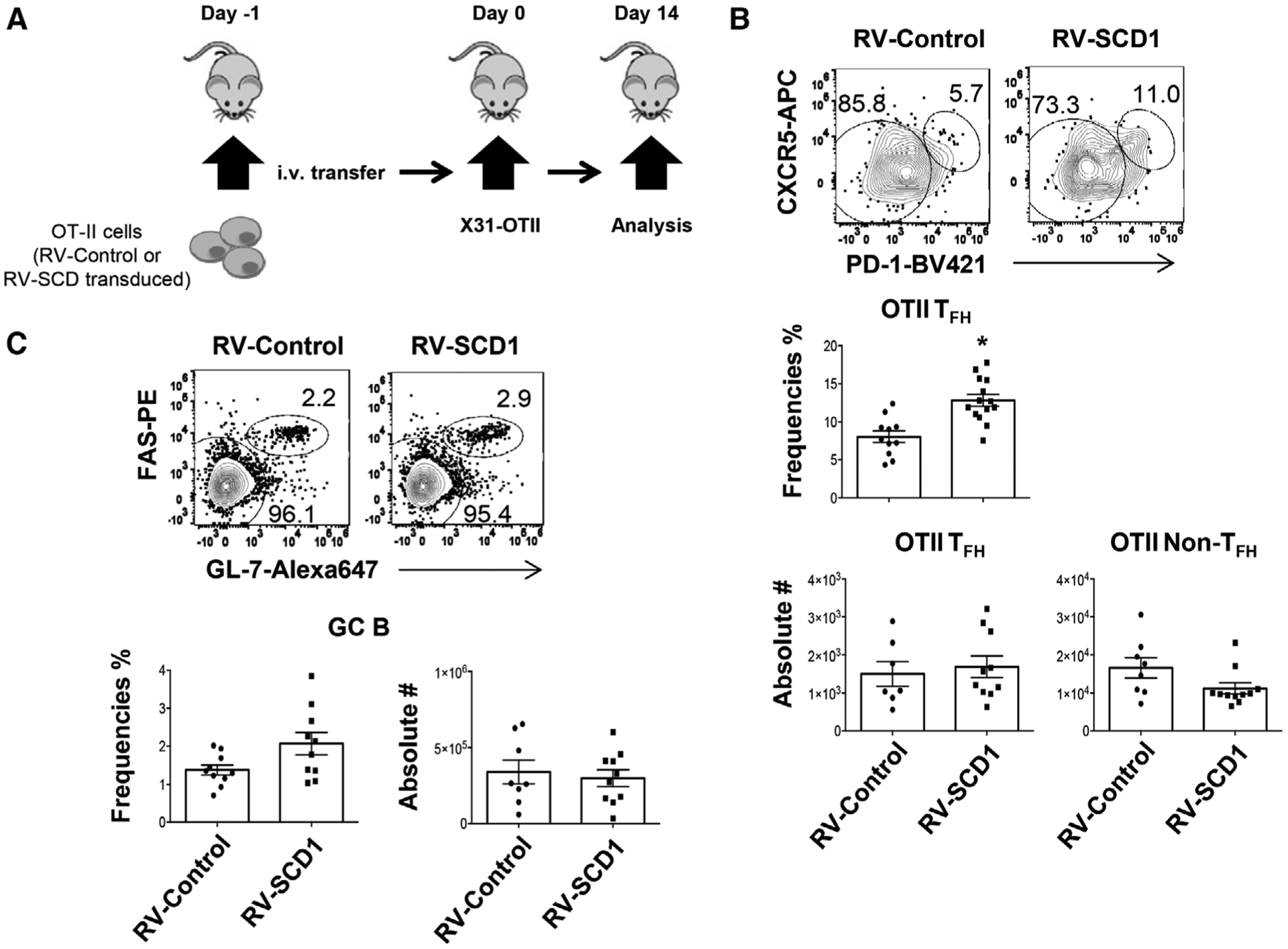
Stearoyl-CoA desaturases (SCD) are endoplasmic reticulum (ER)-associated enzymes that catalyze the synthesis of the monounsaturated fatty acids (MUFAs). As such, SCD play important roles in maintaining the intracellular balance between saturated fatty acid (SFAs) and MUFAs. The roles of SCD in CD4+ T-helper cell responses are currently unexplored. Here, we have found that murine and human follicular helper T (TFH ) cells express higher levels of SCD compared to non-TFH cells. Further, the expression of SCD in TFH cells is dependent on the TFH lineage-specification transcription factor BCL6. We found that the inhibition of SCD impaired TFH cell maintenance and shifted the balance between TFH and follicular regulatory T (TFR ) cells in the spleen. Consequently, SCD inhibition dampened germinal center B-cell responses following influenza immunization. Mechanistically, we found that SCD inhibition led to increased ER stress and enhanced TFH cell apoptosis in vitro and in vivo. These results reveal a possible link between fatty acid metabolism and cellular and humoral responses induced by immunization or potentially, autoimmunity.

Keywords: Endoplasmic reticulum stress; Follicular help T cells; Germinal center B cells; Lipid metabolism; Stearoyl-CoA desaturase 1.


Conflict of interest statement

Conflict of interest: The authors declare no commercial or financial conflict of interest.

Figures

Figure 1.
Figure 1.
SCD inhibition suppresses TFH but not TH1 responses. WT B6 mice were immunized with X31 and treated with indicated inhibitors starting at 4 days postimmunization (d.p.i.). (A) Frequencies and absolute cell numbers of splenic TFH, non-TFH, GC B cells were measured by flow cytometry at 14 d.p.i. (B) Representative dot plot and absolute cell number of splenic CD4+ T or B cells at 14 d.p.i. (C) IFN-γ production by CD4+ or CD8+ T cells were measured through intracellular staining (ICS) following restimulation with NP311–325 (CD4+ T-cell epitope) or NP366–374 (CD8+ T-cell epitope) peptides in vitro at 14 d.p.i. Combined results in A are obtained from three independent experiments (two to five mice per group). Representative data in B and C are obtained from at least two independent experiments (four to five mice per group). Results are given as mean ± SE with one-way ANOVA (A) or mean ± SEM unpaired t-test (B, C). *indicates significant differences (p < 0.05), **p < 0.01, ***p < 0.001, and ****p < 0.0001.
Figure 2.
Figure 2.
Human and mouse TFH express higher levels of SCD mRNA. (A) Relative SCD1 expression in published microarray data (GEO# GSE50391) CD45RO+CXCR5 (non-TFH), CD45RO+CXCR5int (TFH), and CD45RO+CXCR5hi (GC-TFH) cells from human tonsils (five donors per group). (B) Relative expression of Fasn, Scd1, and Scd2 in published microarray data (GEO# GSE40068) from BCL6hiCXCR5+ (TFH) and BCL6CXCR5 (non-TFH) cells of the mice immunized with KLH in CFA (two samples each pooled from three mice per group). (C) Relative expression of Fasn, Scd1, Scd2, and Bcl6 in published RNAseq data (GEO# GSE124883) from LN, spleen, or blood TFH and conventional T cells (Tconv) following NP-OVA immunization (three samples each pooled form ten mice per group). (D) Relative expression of Fasn, Scd1, and Scd2 in sorted splenic TFH (PD-1+CXCR5+) and non-TFH (PD1CXCR5) cells isolated from X31 immunized mice at 14 d.p.i. Representative results of (D) obtained from two independent experiments (five mice per groups). Results are given as mean ± SEM with one- (A) or two- (C) way ANOVA or unpaired t-test (D). *indicates significant differences (p < 0.05), **p < 0.01, ***p < 0.001, and ****p < 0.0001.
Figure 3.
Figure 3.
BCL6 promotes SCD1 expression. (A) Relative expression of Scd1 and Scd2 in WT or BCL6 KO CD4+ T cells under the TFH cell polarizing condition for 4 days. (B) Western blot analysis of SCD1 protein levels in WT or BCL6 KO CD4+ T cells under the TFH cell polarizing condition for 5 days. (C) Scd1, Scd2, and Bcl6 expression in the BCL6-retroviral transduced CD4+ T cells under the TFH cell polarizing condition. (D) WT or BCL6 KO-OTII cells were transfer into CD45.1 congenic mice at one day before X31-OTII virus immunization. After day 14 postimmunization, the OTII cells were sorted and then Scd1 and Scd2 were measured by quantitative RT-PCR (three mice per group in two independent experiments). Representative graphs are obtained from at least two (A, B, and C) independent experiments (two mice per group). Results are given as mean ± SEM with unpaired t-test: *p < 0.05; **p < 0.01; and ***p < 0.001.
Figure 4.
Figure 4.
Inhibition of SCD suppresses TFH and GC B maintenance, but not TFR responses. (A-B) Frequencies and absolute cell number of splenic TFH and GC B cells in the mice immunized with X31 virus and treated with SCD inhibitor at indicated d.p.i. (C) Frequencies and (D) cell numbers of splenic TFR and TFH cells were measured in the mice immunized with X31 virus and treated with SCD inhibitor at 14 d.p.i. (E) The titer of influenza-specific IgG was measured by ELISA from blood serum at 14 d.p.i. Representative data are obtained from at least two independent experiments (two to four mice per group). Results are given as mean ± SE with two-way ANOVA (A and B) or unpaired t-test (C and E): *p < 0.05; and ***p < 0.001.
Figure 5.
Figure 5.
SCD inhibition promotes TFH apoptosis and ER stress gene expression. (A) Caspase 3/7 activity and (B) proliferation (indicated by Ki67 staining) of splenic TFH cells from the mice immunized with X31 virus and treated with SCD inhibitor at 8 and 14 d.p.i. (C) Naïve CD4+ T cells were stimulated under TFH polarization condition and treated with increased doses of SCD inhibitor, active caspase 3/7+ cells were measured at day 4 postactivation. (D) The expression of ER stress-related genes in CD4+ T cells under TFH polarization with or without SCD inhibitor (10 μM) was determined by qRT-PCR. (E) ATF4 protein level in CD4+ T cells under TFH polarization with or without SCD inhibitor. (F) The expression of ER stress-related genes in CD4+ T cells under TFH polarization with or without SCD1 transduction was determined by qRT-PCR. Combine data (A and B) are obtained from two independent experiments (two to five mice per group). Representative results (C and E) are shown from two independent experiments (two mice per group). Results are given as mean ± SEM with unpaired t-test: *p < 0.05.
Figure 6.
Figure 6.
Enforced SCD1 expression in CD4+ T cells promotes TFH responses in vivo. Mock or SCD1-retroviral transduced OTII cells were adoptively transferred into CD45.1 congenic mice. Then the mice were immunized with X31-OTII. (A) Schematics of experimental design. (B) Frequency of TFH cells in OTII cells and cell numbers of TFH and non-TFH cells in CD45.2+ donor cells at 14 d.p.i. (C) Frequency and cell numbers of GC B cells in spleen at 14 d.p.i. Results were pooled from three independent experiments (three to four mice per group). Results are given as mean ± SEM with unpaired t-test: *p < 0.05.

References

    1. Crotty S, T follicular helper cell differentiation, function, and roles in disease. Immunity 2014. 41: 529–542. - PMC - PubMed
    1. Gensous N, Charrier M, Duluc D, Contin-Bordes C, Truchetet ME, Lazaro E, Duffau P et al., T follicular helper cells in autoimmune disorders. Front. Immunol 2018. 9: 1637. - PMC - PubMed
    1. Nurieva RI, Chung Y, Martinez GJ, Yang XO, Tanaka S, Matskevitch TD, Wang YH et al., Bcl6 mediates the development of T follicular helper cells. Science 2009. 325: 1001–1005. - PMC - PubMed
    1. Choi YS, Kageyama R, Eto D, Escobar TC, Johnston RJ, Monticelli L, Lao C et al., ICOS receptor instructs T follicular helper cell versus effector cell differentiation via induction of the transcriptional repressor Bcl6. Immunity 2011. 34: 932–946. - PMC - PubMed
    1. Oestreich KJ, Read KA, Gilbertson SE, Hough KP, McDonald PW, Krishnamoorthy V and Weinmann AS, BCL-6 directly represses the gene program of the glycolysis pathway. Nat. Immunol 2014. 15: 957–964. - PMC - PubMed

Publication types

Substances