. 2020 Jul;177(14):3197-3209.
doi: 10.1111/bph.15042. Epub 2020 Apr 1.

The effect of maslinic acid on cognitive dysfunction induced by cholinergic blockade in mice

Affiliations

The effect of maslinic acid on cognitive dysfunction induced by cholinergic blockade in mice

Ho Jung Bae et al. Br J Pharmacol. 2020 Jul.

Abstract

Background and purpose: Alzheimer's disease (AD) is the most prevalent disease associated with cognitive dysfunction. Current AD therapeutic agents have several gastrointestinal or psychological adverse effects and therefore, novel therapeutic agents with fewer adverse effects must be developed. Previously, we demonstrated that oleanolic acid, which is similar in chemical structure to maslinic acid, ameliorates cognitive impairment through the activation of tropomyosin receptor kinase (TrkB)-ERK-cAMP response element-binding protein (CREB) phosphorylation and increased levels of brain-derived neurotrophic factor (BDNF). In the present study, we investigate the effect of maslinic acid on cholinergic blockade-induced memory impairment in mice.

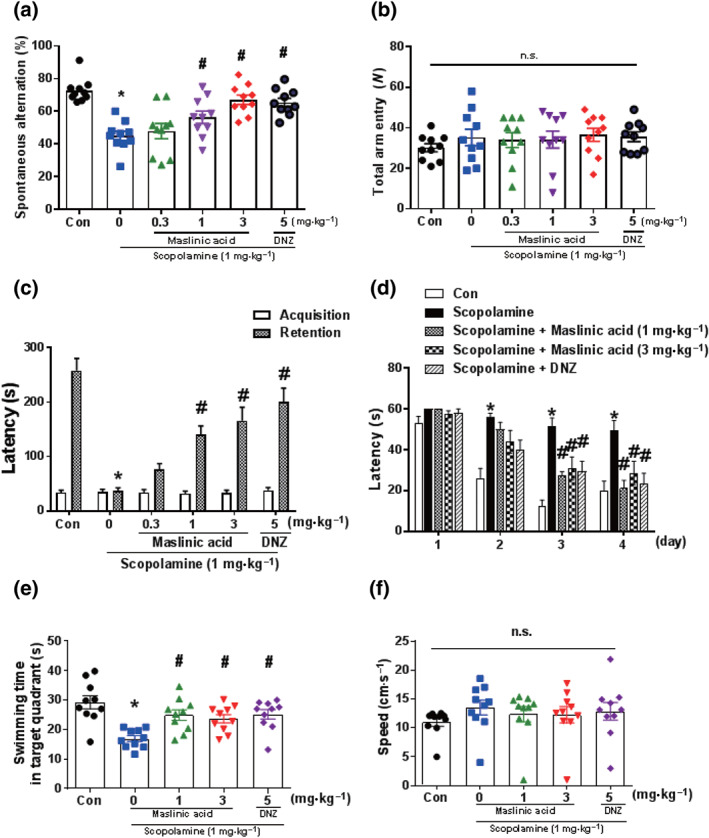
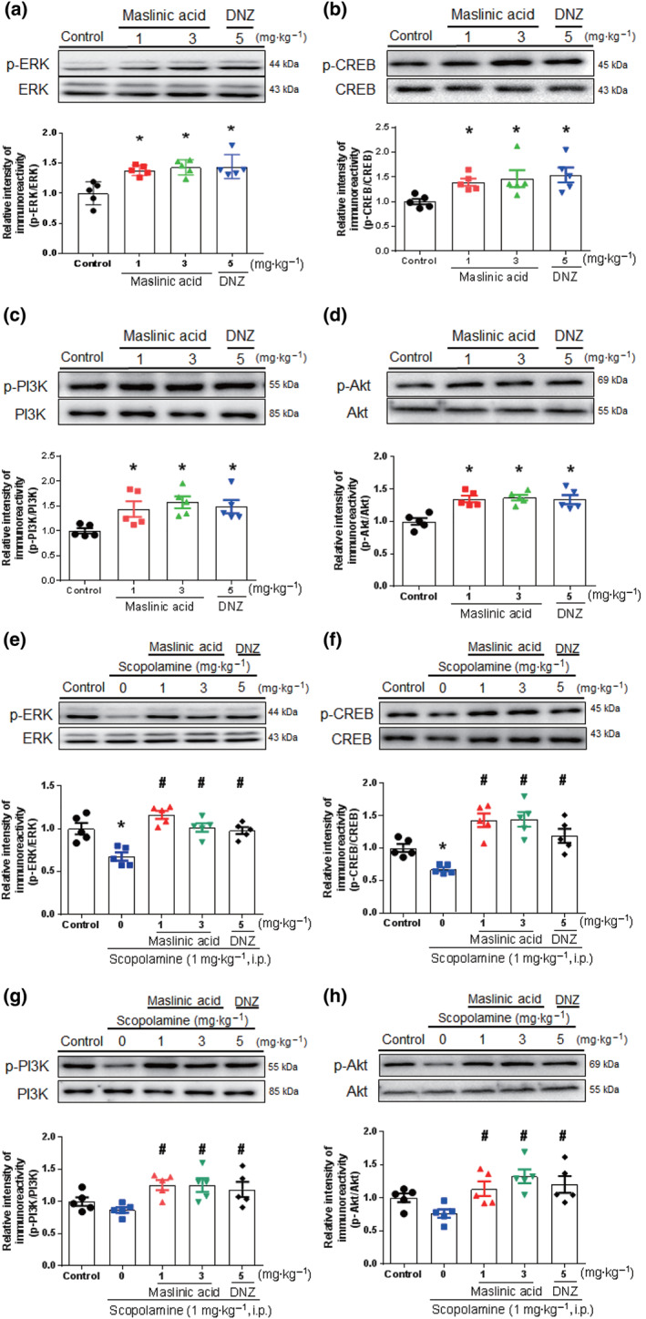
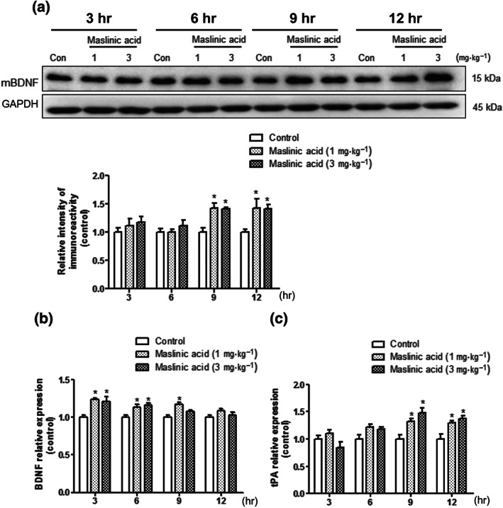
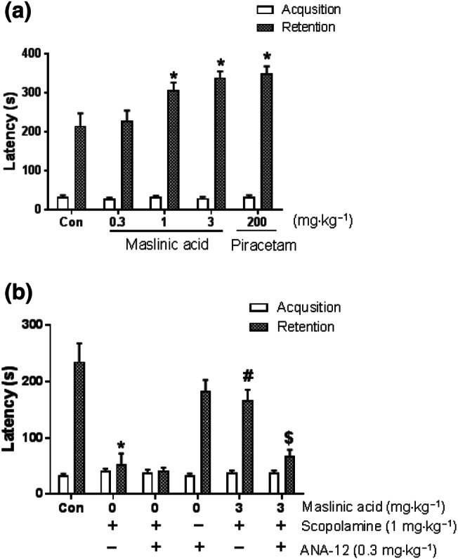
Methods and key results: Maslinic acid reversed scopolamine-induced memory impairment, as determined by the Y-maze, passive avoidance and Morris water maze tests. In addition, we also observed that ERK-CREB, PI3K and PKB (Akt) phosphorylation levels were increased by maslinic acid administration in the mouse hippocampus. Moreover, we determined that the effects of maslinic acid on scopolamine-induced memory impairment in the passive avoidance test were abolished by a specific TrkB receptor antagonist (ANA-12). Additionally, we observed similar temporal changes in the expression levels between BDNF and tissue plasminogen activator in the hippocampus.

Conclusion and implications: These findings suggest that maslinic acid enhances cognitive function through the activation of BDNF and its downstream pathway signalling in the hippocampus and that it might be a potential therapeutic agent for cognitive decline, such as that observed in AD.


Conflict of interest statement

The authors declare no conflicts of interest.

Figures

FIGURE 1
FIGURE 1
Maslinic acid ameliorates cognitive impairment induced by scopolamine treatment in mice. Mice were administered with maslinic acid (0.3, 1 or 3 mg·kg−1, p.o.) or donepezil (DNZ; 5 mg·kg−1, p.o.) 1 hr before the Y‐maze test (a, b). Scopolamine (1 mg·kg−1, i.p.) was treated 30 min before the Y‐maze test to induce memory impairment. Spontaneous alternation behaviour (a) and the number of total arm entries (b) are presented. In addition, mice were treated with maslinic acid (0.3, 1 or 3 mg·kg−1, p.o.) or DNZ (5 mg·kg−1, p.o.) 1 hr before the acquisition trial of the passive avoidance test, whereas scopolamine (1 mg·kg−1, i.p.) was treated 30 min before the acquisition trial to induce memory impairment. Twenty‐four hours after the acquisition trail, the retention trail was conducted for 300 s. The latency time is presented (c). Furthermore, mice were administered with maslinic acid (0.3, 1 or 3 mg·kg−1, p.o.) or DNZ (5 mg·kg−1, p.o.) 1 hr before the first training trial per day for 4 days during the Morris water maze test. Scopolamine (1 mg·kg−1, i.p.) was treated 30 min before the first training trial per day to induce memory impairment (d–f). The escape latencies during the training trial (d), swimming time in the target zone (e) and swimming speed (f) during the probe test after the training trial are presented. Data were showed as means ± SEM (n = 8–10 per group; *P < .05, compared with the vehicle‐administered control [Con] group; # P < .05, compared with the scopolamine‐administered group; n.s., not significant)
FIGURE 2
FIGURE 2
Maslinic acid facilitates LTP induction in the mouse hippocampus. Hippocampal slices were incubated with maslinic acid (1, 10 or 30 μM) for 2 hr. Extracellular field EPSPs (fEPSP) were recorded in Schaffer–collateral–commissural pathway in area CA1. LTP was induced by high‐frequency stimulation (HFS, each 100 Hz, 1 s). Normalized fEPSP slope for maslinic acid 1 μM (a), 10 μM (b) and 30 μM (c) and normalized LTP ratio at 80‐min time point (d) are presented. Data were showed as means ± SEM (n = 7 per group). *P < .05, compared with the vehicle‐administered control group. The number in the parentheses indicates the concentration of maslinic acid (μM)
FIGURE 3
FIGURE 3
The effects of maslinic acid on the phosphorylation level of ERK, CREB, PI3K and Akt in hippocampal tissue. Mice were treated with maslinic acid (1 or 3 mg·kg−1, p.o.), donepezil (DNZ; 5 mg·kg−1, p.o.) or the same volume of vehicle solution 1 hr before the kill. To examine the effect of scopolamine on the signalling molecules, scopolamine was treated 30 min before kill. The levels of p‐ERK/ERK, p‐CREB/CREB, p‐PI3K/PI3K and p‐Akt/Akt were measured in the naïve (a–d) and scopolamine‐induced memory impairment model (e–h). The immunoreactivity of each band was normalized to the control (taken as 1.0). Data were showed as means ± SEM (n = 5 per group; *P < .05, compared with the vehicle‐administered control group; # P < .05, compared with the scopolamine‐administered group; n.s., not significant)
FIGURE 4
FIGURE 4
The effects of maslinic acid on the BDNF and plasminogen activator, tissue type (tPA) expression in the hippocampus. Mice were killed 3, 6, 9 and 12 hr after the administration of maslinic acid (1 or 3 mg·kg−1, p.o.) or the same volume of vehicle solution. The levels of immunoreactivity of BDNF and GAPDH were measured. The graph shows the ration of BDNF/GAPDH normalized to each time point control (a). By using q‐PCR, the mRNA expression levels of BDNF, tPA and GAPDH were measured in the hippocampus. The mRNA expression levels of BDNF (b) and tPA (c) are presented. The mRNA expression levels were normalized by GAPDH. Data were showed as means ± SEM (n = 5 per group; *P < .05, compared with the vehicle‐administered control [Con] group)
FIGURE 5
FIGURE 5
Memory‐enhancing activity of maslinic acid and TrkB activation involved in cognitive function. For the memory enhancement study, mice were treated with maslinic acid (0.3, 1 or 3 mg·kg−1, p.o.) or piracetam (200 mg·kg−1, p.o.) 1 hr before an acquisition trial. Twenty‐four hours after the acquisition trial, the retention trial was performed for 600 s. As piracetam‐treated group, maslinic acid (1 and 3 mg·kg−1, p.o.) enhanced the latency in a dose‐dependent manner (a). In the case of antagonism study, mice were treated with maslinic acid (3 mg·kg−1, p.o.) or the same amount of vehicle solution 1 hr before the acquisition trial. The specific TrkB receptor blocker, ANA‐12 (0.3 mg·kg−1, i.p.), was treated 30 min before the acquisition trial. Consecutively, scopolamine (1 mg·kg−1, i.p.) was treated 3 min after the ANA‐12 treatment. The graph represents the latency of the acquisition and retrieval trials (b). Data were showed as means ± SEM (n = 10 per group; *P < .05, compared with the vehicle‐administered control [Con] group; # P < .05, compared with the scopolamine‐administered group; $ P < .05, compared with the maslinic acid and scopolamine co‐treatment group)

References

    1. Alexander, S. P. H. , Kelly, E. , Mathie, A. , Peters, J. A. , Veale, E. L. , Armstrong, J. F. , … Southan, C. (2019). The Concise Guide to PHARMACOLOGY 2019/20: Introduction and other protein targets. British Journal of Pharmacology, 176(Suppl 1), S1–S20. - PMC - PubMed
    1. Anisman, H. (1975). Dissociation of disinhibitory effects of scopolamine: Strain and task factors. Pharmacology, Biochemistry, and Behavior, 3, 613–618. - PubMed
    1. Bajo, R. , Pusil, S. , Lopez, M. E. , Canuet, L. , Pereda, E. , Osipova, D. , … Pekkonen, E. (2015). Scopolamine effects on functional brain connectivity: A pharmacological model of Alzheimer's disease. Scientific Reports, 5, 9748 10.1038/srep09748 - DOI - PMC - PubMed
    1. Ballatore, C. , Lee, V. M. , & Trojanowski, J. Q. (2007). Tau‐mediated neurodegeneration in Alzheimer's disease and related disorders. Nature Reviews. Neuroscience, 8, 663–672. - PubMed
    1. Bishop, N. A. , Lu, T. , & Yankner, B. A. (2010). Neural mechanisms of ageing and cognitive decline. Nature, 464, 529–535. 10.1038/nature08983 - DOI - PMC - PubMed

Publication types

MeSH terms