Review
. 2020 May;111(5):1452-1460.
doi: 10.1111/cas.14377. Epub 2020 Mar 21.

Artificial intelligence in oncology

Affiliations
Review

Artificial intelligence in oncology

Hideyuki Shimizu et al. Cancer Sci. 2020 May.

Abstract

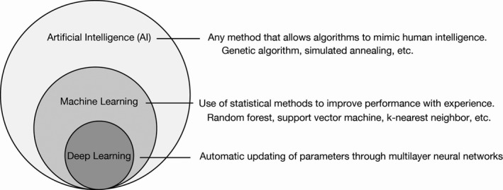
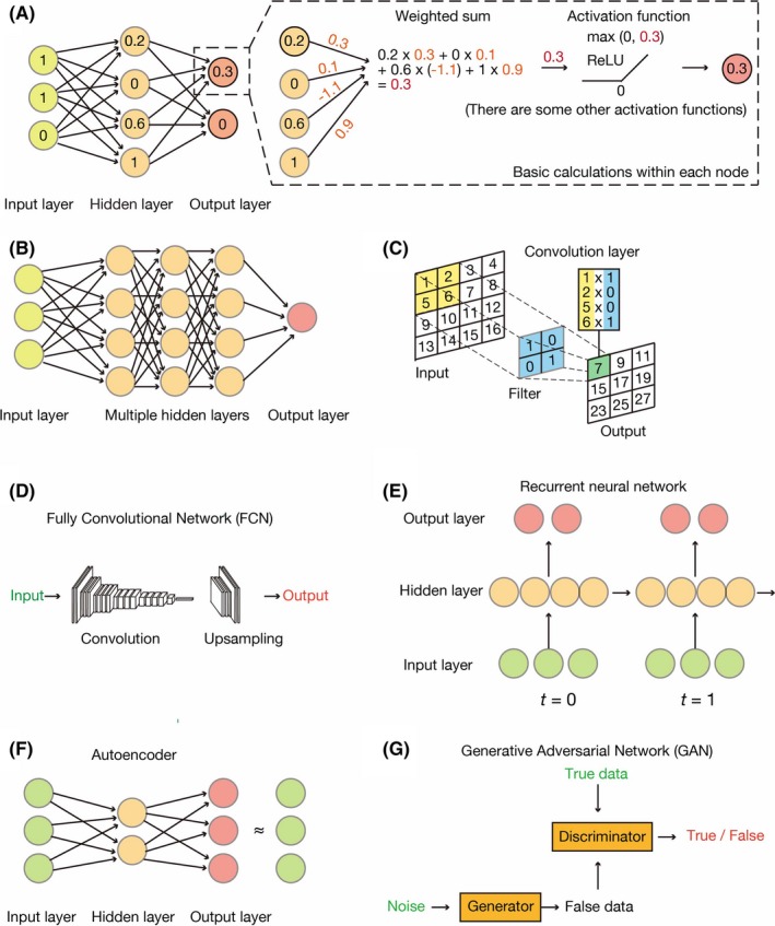
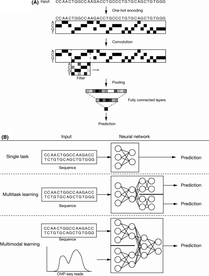
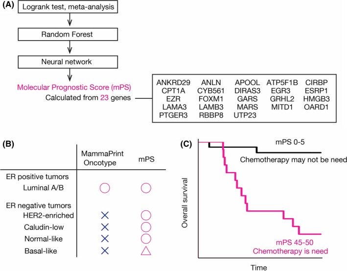
Artificial intelligence (AI) has contributed substantially to the resolution of a variety of biomedical problems, including cancer, over the past decade. Deep learning, a subfield of AI that is highly flexible and supports automatic feature extraction, is increasingly being applied in various areas of both basic and clinical cancer research. In this review, we describe numerous recent examples of the application of AI in oncology, including cases in which deep learning has efficiently solved problems that were previously thought to be unsolvable, and we address obstacles that must be overcome before such application can become more widespread. We also highlight resources and datasets that can help harness the power of AI for cancer research. The development of innovative approaches to and applications of AI will yield important insights in oncology in the coming decade.

Keywords: artificial intelligence; deep learning; machine learning; oncology; personalized medicine.


Conflict of interest statement

The authors declare no potential conflicts of interest.

Figures

Figure 1
Figure 1
Artificial Intelligence (AI), machine learning and deep learning. AI refers to a broad range of computational methods that mimic human intelligence. Machine learning is a subfield of AI that relies on statistical methods to detect hidden patterns within a dataset. Deep learning is a subfield of machine learning that harnesses the power of multilayered networks
Figure 2
Figure 2
Common architectures of neural networks. A, The simplest neural network comprises three layers: an input layer, a hidden layer and an output layer. Each node has some value and transmits its signal to the next layer. It first sums all weighted inputs and then transmits the resulting value to an activation function (rectified linear unit, or ReLU, in this case). B, A typical deep neural network, also known as a dense neural network, has multiple hidden layers, the nodes of each of which calculate values in the same manner as shown in (A). C, A convolutional neural network (CNN) applies multiple convolution layers before feeding the data into a dense neural network. The convolution layers apply filters (or kernels) to grid‐based data. D, A fully convolutional network (FCN) is a variant of a CNN in that it lacks densely connected layers. E, A recurrent neural network (RNN) is a special network designed for time‐series data. Each hidden layer holds certain variables and transmits them to the next time step. F, An autoencoder resembles a dense neural network but is trained to output signals that are identical to the inputs. Such networks offer a means to encode and decode data, with encoded features being stored in the hidden layer. G, A generative adversarial network (GAN) consists of two independent neural networks: a generator and a discriminator. The generator attempts to create new data (false data) that resemble the true data. In contrast, the discriminator discriminates real data from artificial data created by the generator. Alternate training of these two networks helps to decipher the complex rules underlying the data
Figure 3
Figure 3
Deep learning for cancer genomics. A, Sequence data are converted to a binary map (one‐hot encoding) and several filters are applied (1D‐CNN), resulting in the transformation of genomics data into numerical vectors. The remaining procedure is the same as for common tasks (updating of weights to minimize loss). B, A typical workflow receives one type of data and outputs prediction. In multitask learning, multiple types of prediction (such as clinical impact of a mutation and biological activity of a gene) are generated by the shared network and specified networks for each task. Conversely, in multimodal learning, the network integrates different types of information (such as sequence data and chromatin accessibility) and outputs prediction
Figure 4
Figure 4
Prediction of prognosis of breast cancer patients. A, All human protein‐coding genes were evaluated using a series of methods (log‐rank test, meta‐analysis, machine learning with the random forest method and a neural network). The molecular Prognostic Score (mPS) based on 23 prognostic genes predicts the prognosis of breast cancer patients. B, mPS stratifies not only estrogen receptor (ER)‐positive but also ER‐negative patients, in contrast to existing methods such as MammaPrint and Oncotype. C, Chemotherapy may not be necessary for patients with a low mPS because their prognosis is fairly good

Similar articles

Cited by

References

    1. Rajkomar A, Dean J, Kohane I. Machine learning in medicine. N Engl J Med. 2019;380:1347‐1358. - PubMed
    1. LeCun Y, Bengio Y, Hinton G. Deep learning. Nature. 2015;521:436‐444. - PubMed
    1. Teare P, Fishman M, Benzaquen O, Toledano E, Elnekave E. Malignancy detection on mammography using dual deep convolutional neural networks and genetically discovered false color input enhancement. J Digit Imaging. 2017;30:499‐505. - PMC - PubMed
    1. Ehteshami Bejnordi B, Veta M, Johannes van Diest P, et al. Diagnostic assessment of deep learning algorithms for detection of lymph node metastases in women with breast cancer. JAMA. 2017;318:2199‐2210. - PMC - PubMed
    1. Gulshan V, Peng L, Coram M, et al. Development and validation of a deep learning algorithm for detection of diabetic retinopathy in retinal fundus photographs. JAMA. 2016;316:2402‐2410. - PubMed