

Non-clinical efficacy, safety and stable clinical cell processing of induced pluripotent stem cell-derived anti-glypican-3 chimeric antigen receptor-expressing natural killer/innate lymphoid cells
- PMID: 32133731
- PMCID: PMC7226201
- DOI: 10.1111/cas.14374
Non-clinical efficacy, safety and stable clinical cell processing of induced pluripotent stem cell-derived anti-glypican-3 chimeric antigen receptor-expressing natural killer/innate lymphoid cells
Abstract
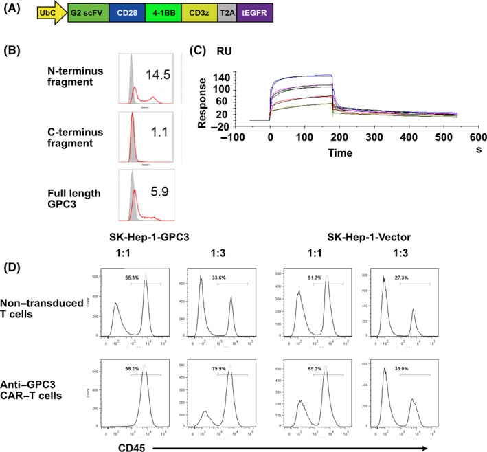
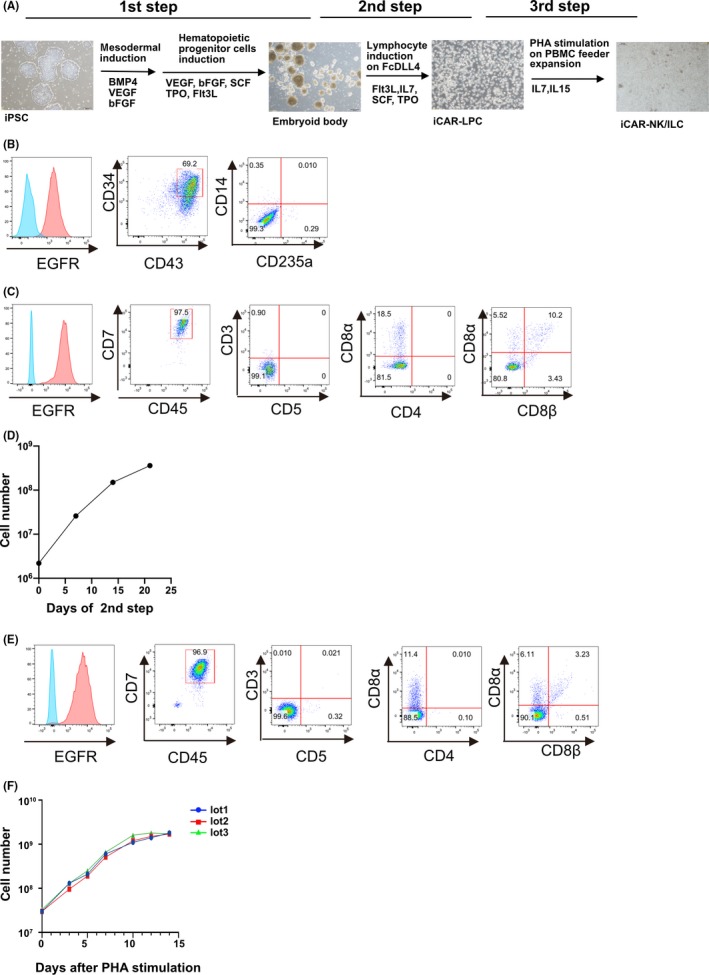
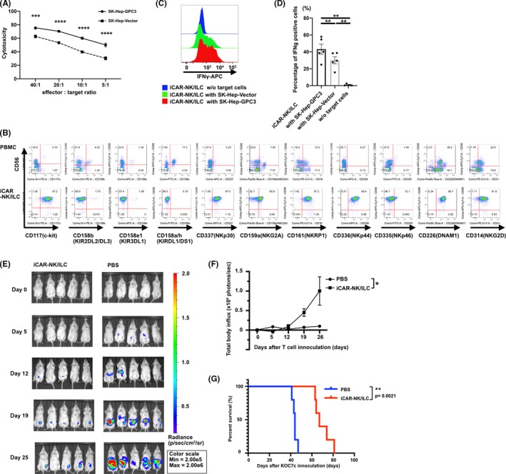
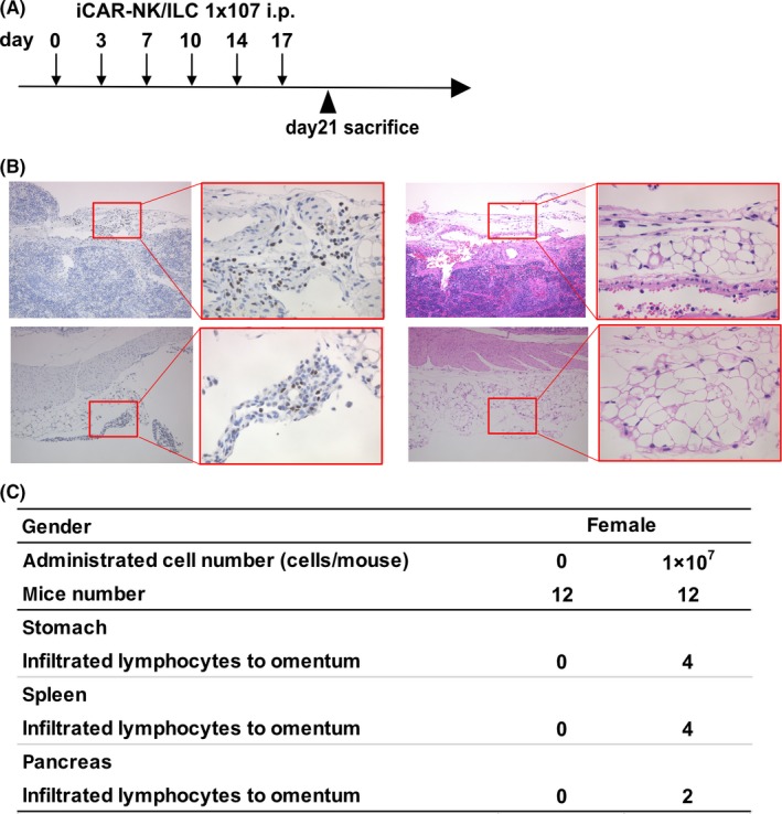
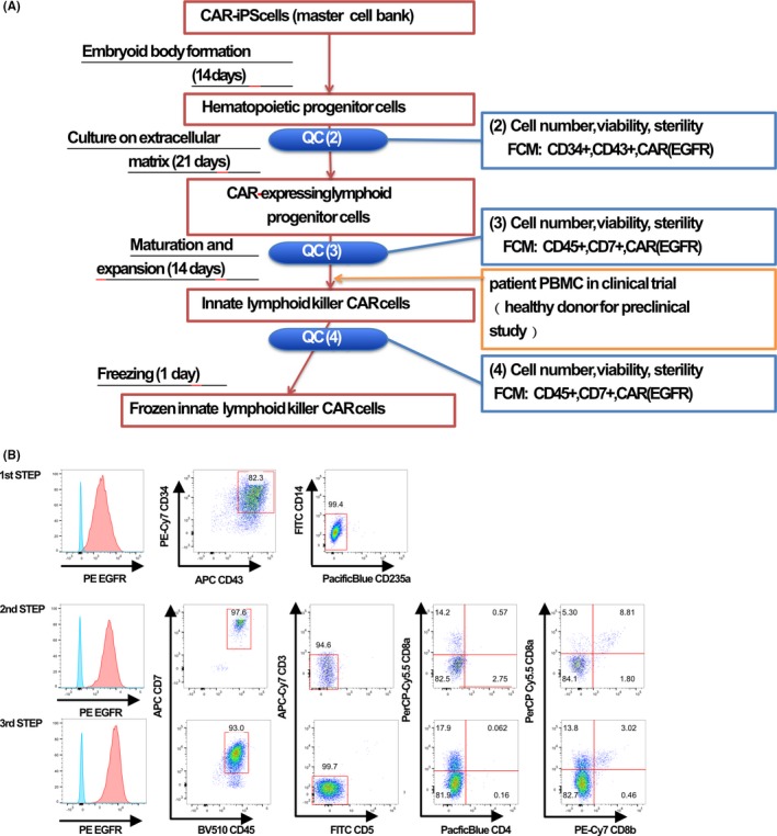
The use of allogeneic, pluripotent stem-cell-derived immune cells for cancer immunotherapy has been the subject of recent clinical trials. In Japan, investigator-initiated clinical trials will soon begin for ovarian cancer treatment using human leukocyte antigen (HLA)-homozygous-induced pluripotent stem cell (iPSC)-derived anti-glypican-3 (GPC3) chimeric antigen receptor (CAR)-expressing natural killer/innate lymphoid cells (NK/ILC). Using pluripotent stem cells as the source for allogeneic immune cells facilitates stringent quality control of the final product, in terms of efficacy, safety and producibility. In this paper, we describe our methods for the stable, feeder-free production of CAR-expressing NK/ILC cells from CAR-transduced iPSC with clinically relevant scale and materials. The average number of cells that could be differentiated from 1.8-3.6 × 10⁶ iPSC within 7 weeks was 1.8-4.0 × 10⁹ . These cells showed stable CD45/CD7/CAR expression, effector functions of cytotoxicity and interferon gamma (IFN-γ) production against GPC3-expressing tumor cells. When the CAR-NK/ILC cells were injected into a GPC3-positive, ovarian-tumor-bearing, immunodeficient mouse model, we observed a significant therapeutic effect that prolonged the survival of the animals. When the cells were injected into immunodeficient mice during non-clinical safety tests, no acute systemic toxicity or tumorigenicity of the final product or residual iPSC was observed. In addition, our test results for the CAR-NK/ILC cells generated with clinical manufacturing standards are encouraging, and these methods should accelerate the development of allogeneic pluripotent stem cell-based immune cell cancer therapies.
Keywords: GPC3; ILC; NK; chimeric antigen receptor; iPSC; immunotherapy.
© 2020 The Authors. Cancer Science published by John Wiley & Sons Australia, Ltd on behalf of Japanese Cancer Association.
Conflict of interest statement
Shin Kaneko is a founder, shareholder and chief scientific officer at Thyas and received research funding from Takeda Pharmaceutical, Kirin Holdings, Terumo, Tosoh, Sumitomo Chemical and Thyas. Tetsuya Nakatsura is a shareholder at Killer T Save You. Koji Tamada is a shareholder of Noile‐Immune Biotech and receives consulting fees and research funding from Noile‐Immune Biotech. Yutaka Yasui is an employee of Thyas. The remaining authors declare no competing financial interests.
Figures
References
-
- Kiessling R, Klein E, Wigzell H. Natural killer cells in the mouse. Eur J Immunol. 1975;5:112‐117. - PubMed
-
- Spits H, Artis D, Colonna M, et al. Innate lymphoid cells‐a proposal for uniform nomenclature. Nat Rev Immunol. 2013;13:145‐149. - PubMed
-
- Chang YH, Connolly J, Shimasaki N, Mimura K, Kono K, Campana D. A chimeric receptor with NKG2D specificity enhances natural killer cell activation and killing of tumor cells. Cancer Res. 2013;73:1777‐1786. - PubMed
MeSH terms
Substances
Grants and funding
- 15H04655/the Ministry of Education, Culture, Sports, Science and Technology Japan
- 25293226/the Ministry of Education, Culture, Sports, Science and Technology Japan
- 26293357/the Ministry of Education, Culture, Sports, Science and Technology Japan
- Japan Agency for Medical Research and Development
- National Cancer Center Research Fund
LinkOut - more resources
Full Text Sources
Other Literature Sources
Research Materials
Miscellaneous
