Review
. 2020 Mar;16(3):e9232.
doi: 10.15252/msb.20199232.

Thermal proteome profiling for interrogating protein interactions

Affiliations
Review

Thermal proteome profiling for interrogating protein interactions

André Mateus et al. Mol Syst Biol. 2020 Mar.

Abstract

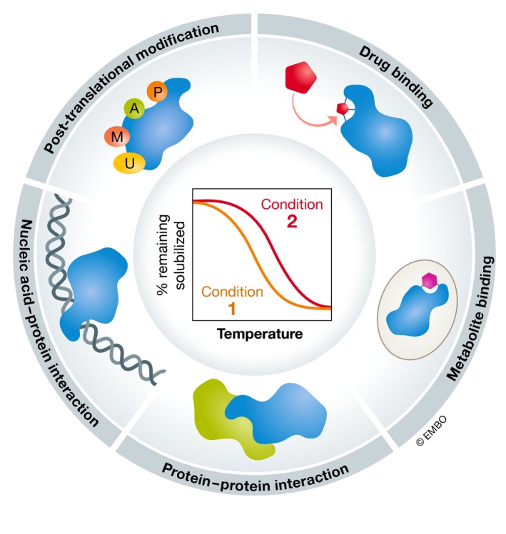
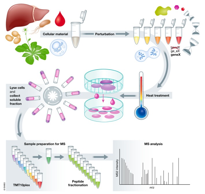
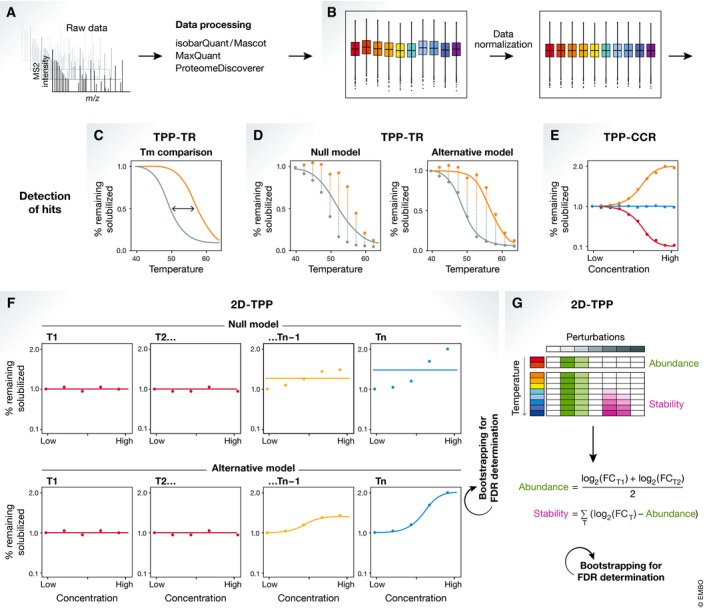
Thermal proteome profiling (TPP) is based on the principle that, when subjected to heat, proteins denature and become insoluble. Proteins can change their thermal stability upon interactions with small molecules (such as drugs or metabolites), nucleic acids or other proteins, or upon post-translational modifications. TPP uses multiplexed quantitative mass spectrometry-based proteomics to monitor the melting profile of thousands of expressed proteins. Importantly, this approach can be performed in vitro, in situ, or in vivo. It has been successfully applied to identify targets and off-targets of drugs, or to study protein-metabolite and protein-protein interactions. Therefore, TPP provides a unique insight into protein state and interactions in their native context and at a proteome-wide level, allowing to study basic biological processes and their underlying mechanisms.

Keywords: drug discovery; metabolites; protein complexes; proteomics; thermal proteome profiling.


Conflict of interest statement

The authors declare that they have no conflict of interest.

Figures

Figure 1
Figure 1. Thermal proteome profiling (TPP) provides proteome‐wide information on protein states and interactions
TPP combines the principles of the cellular thermal shift assay (CETSA) with multiplexed quantitative mass spectrometry‐based proteomics. CETSA is based on the principle that proteins denature and become insoluble when subjected to heat. By monitoring the remaining soluble fraction at multiple temperatures, melting profiles for each detected protein can be obtained. The melting profile depends on the context of the protein and can be altered by interactions with small molecules (such as drugs or metabolites), nucleic acids, or other proteins, or post‐translational modifications. CETSA and TPP can be applied in vitro, in situ, and in vivo.
Figure 2
Figure 2. Thermal proteome profiling (TPP) experimental setup
(A) TPP starts by the choice of cellular material to study: cell extracts, intact cells, tissues, or biological fluids, from any domain of life (Archaea, Bacteria, or Eukarya, the latter including Protista, Fungi, Plantae, or Animalia). (B) A perturbation can then be induced: commonly a chemical (e.g., drug or metabolite), genetic (e.g., gene knock‐out or overexpression, or point mutation in a gene), environmental, or enzymatic perturbation. Some of these can be applied in a dose‐ or time‐dependent manner. (C) Samples are then subjected to a short heat treatment to induce protein aggregation. (D) The remaining soluble fraction at each temperature is collected after ultracentrifugation or using multi‐well filter plates. (E) Samples are processed using a bottom‐up proteomics workflow, generally using isobaric tandem mass tags (TMT). Labeled peptides are combined and fractionated. (f) Peptides are analyzed by mass spectrometry.
Figure 3
Figure 3. Thermal proteome profiling (TPP) data analysis
(A) Raw mass spectrometry data are processed to identify and quantify the measured proteins. (B) Data are then normalized to remove any artifacts introduced during the experimental procedure (e.g., different amounts of protein in each sample). Depending on the type of experiment performed, different analysis strategies exist as follows: (C, D) For TPP‐TR experiments, (C) melting points or (D) whole melting profiles can be compared between conditions. (E) For TPP‐CCR, dose–response curves are fit and targets are selected if a certain degree of stabilization and a good coefficient of determination are obtained. (F) For 2D‐TPP experiments with a dose‐dependent setup, a null model (linear) can be compared to an alternative model (sigmoidal) by comparing the goodness of fit of both models. The false discovery rate (FDR) is inferred by using a bootstrapping approach. (G) For 2D‐TPP with discrete perturbations, a reference condition is selected and fold changes for all other conditions are calculated. To separate abundance from thermal stability effects, this method integrates the relative log‐transformed fold changes measured at the first two temperatures, which are assumed to solely reflect abundance changes. Then, the log‐transformed fold changes are adjusted for the abundance effect and the integral of the log‐transformed fold changes at all temperatures is calculated, which reflect thermal stability changes. In this way, individual perturbations are assigned an abundance and thermal stability score which both are tested for significant deviation from zero by a bootstrapping approach.

References

    1. Aebersold R, Mann M (2016) Mass‐spectrometric exploration of proteome structure and function. Nature 537: 347–355 - PubMed
    1. Aguzzi A, Altmeyer M (2016) Phase separation: linking cellular compartmentalization to disease. Trends Cell Biol 26: 547–558 - PubMed
    1. Azimi A, Caramuta S, Seashore‐Ludlow B, Bostrom J, Robinson JL, Edfors F, Tuominen R, Kemper K, Krijgsman O, Peeper DS et al (2018) Targeting CDK2 overcomes melanoma resistance against BRAF and Hsp90 inhibitors. Mol Syst Biol 14: e7858 - PMC - PubMed
    1. Banani SF, Lee HO, Hyman AA, Rosen MK (2017) Biomolecular condensates: organizers of cellular biochemistry. Nat Rev Mol Cell Biol 18: 285–298 - PMC - PubMed
    1. Banzhaf M, Yau HCL, Verheul J, Lodge A, Kritikos G, Mateus A, Hov AK, Stein F, Wartel M, Pazos M et al (2020) Outer membrane lipoprotein NlpI scaffolds peptidoglycan hydrolases within multi‐enzyme complexes in Escherichia coli. EMBO J 10.15252/embj.2019102246 - DOI - PMC - PubMed

Publication types